Assessing the role of selenium in Minamata disease through reanalysis of historical samples
IF 9.7
1区 环境科学与生态学
Q1 ENVIRONMENTAL SCIENCES
引用次数: 0
Abstract
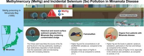
Minamata disease, a severe neurological disorder identified in Japan in 1956, results from methylmercury (MeHg) intoxication in humans due to environmental contamination. Before MeHg was recognized as the cause, selenium (Se) was suspected of being the potential cause owing to elevated Se levels in patients’ organs. Subsequent animal studies indicated that Se mitigates MeHg toxicity; however, its role in Minamata disease remains unexplored. We analyzed Hg and Se in historical samples of the industrial wastes (n = 4) on the factory site, sediments (n = 9), and fish/shellfish (n = 16) in Minamata Bay, and organs of patients with Minamata disease (n = 12). All samples showed elevated levels of both Hg and Se, providing the first evidence that Se was also discharged into Minamata Bay, entering the food chain and accumulating at high levels in patient organs. The Hg/Se molar ratio in contaminated shellfish (median > 3.0) indicated exceptionally high MeHg exposure, far exceeding the ordinary level (< 1.0). Patients exhibited significantly increased Se levels in the liver and kidney but lower amounts in the brain. Notably, median Hg/Se molar ratios exceeding 4.0 were observed, particularly in the cerebrum and cerebellum in acute cases, closely mirroring the molar ratios found in seafood. The elevated Hg/Se molar ratio in the brain helps explain the severe neurological damage in patients’ central nervous systems, despite higher Hg levels in the liver and kidney compared to the brain. These findings provide important insight into the mechanism of MeHg intoxication and highlight the risks associated with MeHg-contaminated seafood, aiding efforts to protect consumers.


通过重新分析历史样本评估硒在水俣病中的作用
水俣病是1956年在日本发现的一种严重的神经系统疾病,是由于环境污染导致人类甲基汞中毒造成的。在MeHg被确认为病因之前,由于患者器官中的硒水平升高,人们怀疑硒(Se)是潜在的病因。随后的动物研究表明,硒可以减轻甲基汞的毒性;然而,它在水俣病中的作用仍未被探索。我们分析了工厂厂区工业废弃物(n = 4)、水俣湾沉积物(n = 9)、鱼/贝类(n = 16)和水俣病患者器官(n = 12)的历史样品中汞和硒的含量。所有样本均显示汞和硒水平升高,首次提供证据表明,硒也被排放到水俣湾,进入食物链并在患者器官中大量积累。受污染贝类的汞硒摩尔比(中位数 >; 3.0)表明甲基汞暴露异常高,远远超过正常水平(<;1.0)。患者肝脏和肾脏中的硒含量明显增加,但大脑中的硒含量较低。值得注意的是,汞/硒的摩尔比中值超过4.0,特别是在急性病例的大脑和小脑中,与海鲜中的摩尔比密切相关。大脑中汞/硒摩尔比的升高有助于解释患者中枢神经系统的严重神经损伤,尽管肝脏和肾脏中的汞含量高于大脑。这些发现为甲基汞中毒的机制提供了重要的见解,并强调了与甲基汞污染的海产品相关的风险,有助于保护消费者。
本文章由计算机程序翻译,如有差异,请以英文原文为准。
求助全文
约1分钟内获得全文
求助全文
来源期刊

Environment International
环境科学-环境科学
CiteScore
21.90
自引率
3.40%
发文量
734
审稿时长
2.8 months
期刊介绍:
Environmental Health publishes manuscripts focusing on critical aspects of environmental and occupational medicine, including studies in toxicology and epidemiology, to illuminate the human health implications of exposure to environmental hazards. The journal adopts an open-access model and practices open peer review.
It caters to scientists and practitioners across all environmental science domains, directly or indirectly impacting human health and well-being. With a commitment to enhancing the prevention of environmentally-related health risks, Environmental Health serves as a public health journal for the community and scientists engaged in matters of public health significance concerning the environment.
 求助内容:
求助内容: 应助结果提醒方式:


