Maciej Biały, Wacław Marceli Adamczyk, Tomasz Stranc, Grzegorz Szlachta, Rafał Gnat
{"title":"M-mode ultrasound evaluation of lateral abdominal muscle postural response to load: an exploratory study.","authors":"Maciej Biały, Wacław Marceli Adamczyk, Tomasz Stranc, Grzegorz Szlachta, Rafał Gnat","doi":"10.15557/jou.2024.0037","DOIUrl":null,"url":null,"abstract":"<p><strong>Aim: </strong>There is a need to evaluate the tissue deformation index of lateral abdominal muscles using M-mode ultrasound in a cohort of healthy subjects to establish a convenient reference point for clinical reasoning in patients. The aim of the study was to assess differences in the tissue deformation index between individual lateral abdominal muscles regardless of body side, compare these differences in the tissue deformation index on the right and left sides of the body, and evaluate side-to-side differences in the tissue deformation index within individual lateral abdominal muscles.</p><p><strong>Material and methods: </strong>In a group of 126 healthy volunteers (59 females), the postural response of lateral abdominal muscles to external perturbation in the form of rapid arm abduction with load was recorded on both sides of the body, and the tissue deformation index was calculated.</p><p><strong>Results: </strong>The mean values of the tissue deformation index form an increasing gradient from deep to superficial lateral abdominal muscles: 0.06%/ms for the transversus abdominis, 0.084%/ms for the internal oblique and 0.151%/ms for the external oblique (<i>p</i> <0.001). Side-to-side intra-muscle differences were significant only for the transverse abdominis (right: 0.047%/ms; left: 0.070%; <i>p</i> <0.01).</p><p><strong>Conclusions: </strong>The tissue deformation index values differ significantly among individual lateral abdominal muscles and form a characteristic gradient: transversus abdominis < internal oblique < external oblique. The transversus abdominis muscle shows significant asymmetry in the tissue deformation index between the left and right sides of the body.</p>","PeriodicalId":45612,"journal":{"name":"Journal of Ultrasonography","volume":"24 99","pages":"1-7"},"PeriodicalIF":1.5000,"publicationDate":"2024-12-23","publicationTypes":"Journal Article","fieldsOfStudy":null,"isOpenAccess":false,"openAccessPdf":"https://www.ncbi.nlm.nih.gov/pmc/articles/PMC11665055/pdf/","citationCount":"0","resultStr":null,"platform":"Semanticscholar","paperid":null,"PeriodicalName":"Journal of Ultrasonography","FirstCategoryId":"1085","ListUrlMain":"https://doi.org/10.15557/jou.2024.0037","RegionNum":0,"RegionCategory":null,"ArticlePicture":[],"TitleCN":null,"AbstractTextCN":null,"PMCID":null,"EPubDate":"2024/12/1 0:00:00","PubModel":"eCollection","JCR":"Q3","JCRName":"RADIOLOGY, NUCLEAR MEDICINE & MEDICAL IMAGING","Score":null,"Total":0}
引用次数: 0
Abstract
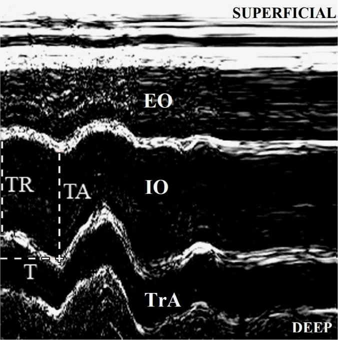
Aim: There is a need to evaluate the tissue deformation index of lateral abdominal muscles using M-mode ultrasound in a cohort of healthy subjects to establish a convenient reference point for clinical reasoning in patients. The aim of the study was to assess differences in the tissue deformation index between individual lateral abdominal muscles regardless of body side, compare these differences in the tissue deformation index on the right and left sides of the body, and evaluate side-to-side differences in the tissue deformation index within individual lateral abdominal muscles.
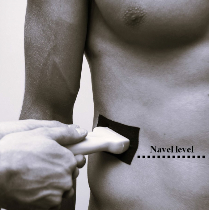
Material and methods: In a group of 126 healthy volunteers (59 females), the postural response of lateral abdominal muscles to external perturbation in the form of rapid arm abduction with load was recorded on both sides of the body, and the tissue deformation index was calculated.
Results: The mean values of the tissue deformation index form an increasing gradient from deep to superficial lateral abdominal muscles: 0.06%/ms for the transversus abdominis, 0.084%/ms for the internal oblique and 0.151%/ms for the external oblique (p <0.001). Side-to-side intra-muscle differences were significant only for the transverse abdominis (right: 0.047%/ms; left: 0.070%; p <0.01).
Conclusions: The tissue deformation index values differ significantly among individual lateral abdominal muscles and form a characteristic gradient: transversus abdominis < internal oblique < external oblique. The transversus abdominis muscle shows significant asymmetry in the tissue deformation index between the left and right sides of the body.


 求助内容:
求助内容: 应助结果提醒方式:


