Tieci Yi, Difei Lu, Yonggang Cui, Zheng Zhang, Xing Yang, Jianhua Zhang, Lin Qiu, Haoyu Weng, Lin Liu, Xiaojiang Duan, Guangyu Zhao, Wei Ma, Ying Gao, Yan Fan
{"title":"<sup>68</sup>Ga-pentixafor PET/CT Is a Supplementary Method for Primary Aldosteronism Subtyping Compared with Adrenal Vein Sampling.","authors":"Tieci Yi, Difei Lu, Yonggang Cui, Zheng Zhang, Xing Yang, Jianhua Zhang, Lin Qiu, Haoyu Weng, Lin Liu, Xiaojiang Duan, Guangyu Zhao, Wei Ma, Ying Gao, Yan Fan","doi":"10.1007/s11307-024-01976-0","DOIUrl":null,"url":null,"abstract":"<p><strong>Purpose: </strong>To investigate the diagnostic efficacy of <sup>68</sup>Ga-pentixafor positron emission tomography/computed tomography (PET/CT) in primary aldosteronism (PA) subtyping and lateralization of aldosterone secretion in PA patients.</p><p><strong>Procedures: </strong>37 patients who were diagnosed with PA, were prospectively enrolled in the study, and underwent adrenal vein sampling (AVS) after <sup>68</sup>Ga-pentixafor PET/CT was conducted. Lateralization index (LI), defined as aldosterone/cortisol ratio in the dominant side to the contralateral adrenal vein when bilateral adrenal vein catheterization succeeded, and the aldosterone/cortisol ratio in the left adrenal vein to IVC (LAV/IVC) when the catheterization of right adrenal vein failed, were applied to determine lateralization side. Statistical analysis was performed using SPSS 21.0.</p><p><strong>Results: </strong>The female proportion of all patients with PA was 32.4% (12/37), and the mean age was 51.3 ± 10.9 years. Patients with bilateral adrenal mass accounted for 54.1% (20/37), and 10 of them (27.0%) had adrenal hyperplasia or adrenal nodules ≤ 1.0 cm. In all 37 patients, the sensitivity, specificity and accuracy of <sup>68</sup>Ga-pentixafor PET/CT in distinguishing lateralization by visualization were 89.3%, 77.8% and 86.5%, respectively. The area under the ROC curve for detecting positive lateralization based on the value of <sup>68</sup>Ga-pentixafor SUV<sub>max</sub> was 0.750 (95%CI 0.578-0.922, p = 0.026). The optimum SUV<sub>max</sub> cut-off value was 6.86, with the sensitivity of 78.6%, specificity of 66.7%, and accuracy of 78.4%. Defining SUV ratio as SUV<sub>max</sub>/SUV of contralateral adrenal gland, the area under the ROC curve for identifying lateralization based on the SUV ratio was 0.710 (95%CI 0.500-0.921, p = 0.061). The optimum SUV ratio cut-off was 2.40, with the sensitivity of 60.7%, specificity of 88.9%, and accuracy of 67.6%. The consistency of <sup>68</sup>Ga-pentixafor PET/CT with AVS was of no significant difference between patients with bilateral adrenal lesions (80.0%, 16/20) and unilateral lesion (94.1%, 16/17; p = 0.737), and no significance was revealed in the consistency between patients with adrenal hyperplasia or adrenal lesion of diameter ≤ 1 cm (81.8%, 9/11) and those with adrenal lesions > 1 cm (88.5%, 23/26; p = 0.884).</p><p><strong>Conclusions: </strong><sup>68</sup>Ga-pentixafor PET/CT showed at least 80% consistency for the lateralization in patients with PA compared with AVS, even in those presented with bilateral adrenal hyperplasia. Visual analysis exhibited better diagnostic efficacy compared with SUV<sub>max</sub> or SUV<sub>max</sub>/SUV of the contralateral adrenal gland.( ChiCTR2300073049. Registered 30 June 2023. Retrospectively registered).</p>","PeriodicalId":18760,"journal":{"name":"Molecular Imaging and Biology","volume":" ","pages":"142-150"},"PeriodicalIF":2.5000,"publicationDate":"2025-02-01","publicationTypes":"Journal Article","fieldsOfStudy":null,"isOpenAccess":false,"openAccessPdf":"https://www.ncbi.nlm.nih.gov/pmc/articles/PMC11805762/pdf/","citationCount":"0","resultStr":null,"platform":"Semanticscholar","paperid":null,"PeriodicalName":"Molecular Imaging and Biology","FirstCategoryId":"3","ListUrlMain":"https://doi.org/10.1007/s11307-024-01976-0","RegionNum":4,"RegionCategory":"医学","ArticlePicture":[],"TitleCN":null,"AbstractTextCN":null,"PMCID":null,"EPubDate":"2024/12/23 0:00:00","PubModel":"Epub","JCR":"Q2","JCRName":"RADIOLOGY, NUCLEAR MEDICINE & MEDICAL IMAGING","Score":null,"Total":0}
引用次数: 0
Abstract
Purpose: To investigate the diagnostic efficacy of 68Ga-pentixafor positron emission tomography/computed tomography (PET/CT) in primary aldosteronism (PA) subtyping and lateralization of aldosterone secretion in PA patients.
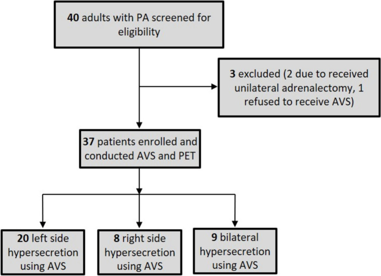
Procedures: 37 patients who were diagnosed with PA, were prospectively enrolled in the study, and underwent adrenal vein sampling (AVS) after 68Ga-pentixafor PET/CT was conducted. Lateralization index (LI), defined as aldosterone/cortisol ratio in the dominant side to the contralateral adrenal vein when bilateral adrenal vein catheterization succeeded, and the aldosterone/cortisol ratio in the left adrenal vein to IVC (LAV/IVC) when the catheterization of right adrenal vein failed, were applied to determine lateralization side. Statistical analysis was performed using SPSS 21.0.
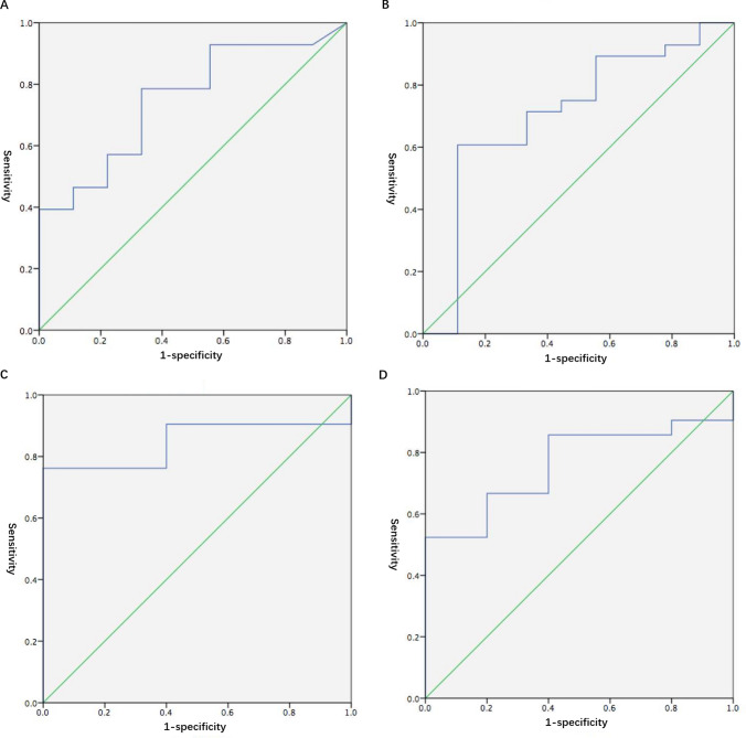
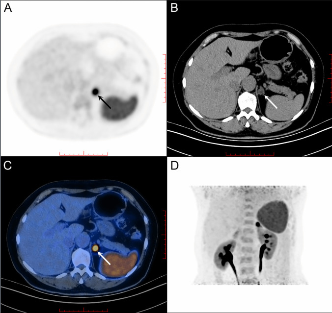
Results: The female proportion of all patients with PA was 32.4% (12/37), and the mean age was 51.3 ± 10.9 years. Patients with bilateral adrenal mass accounted for 54.1% (20/37), and 10 of them (27.0%) had adrenal hyperplasia or adrenal nodules ≤ 1.0 cm. In all 37 patients, the sensitivity, specificity and accuracy of 68Ga-pentixafor PET/CT in distinguishing lateralization by visualization were 89.3%, 77.8% and 86.5%, respectively. The area under the ROC curve for detecting positive lateralization based on the value of 68Ga-pentixafor SUVmax was 0.750 (95%CI 0.578-0.922, p = 0.026). The optimum SUVmax cut-off value was 6.86, with the sensitivity of 78.6%, specificity of 66.7%, and accuracy of 78.4%. Defining SUV ratio as SUVmax/SUV of contralateral adrenal gland, the area under the ROC curve for identifying lateralization based on the SUV ratio was 0.710 (95%CI 0.500-0.921, p = 0.061). The optimum SUV ratio cut-off was 2.40, with the sensitivity of 60.7%, specificity of 88.9%, and accuracy of 67.6%. The consistency of 68Ga-pentixafor PET/CT with AVS was of no significant difference between patients with bilateral adrenal lesions (80.0%, 16/20) and unilateral lesion (94.1%, 16/17; p = 0.737), and no significance was revealed in the consistency between patients with adrenal hyperplasia or adrenal lesion of diameter ≤ 1 cm (81.8%, 9/11) and those with adrenal lesions > 1 cm (88.5%, 23/26; p = 0.884).
Conclusions: 68Ga-pentixafor PET/CT showed at least 80% consistency for the lateralization in patients with PA compared with AVS, even in those presented with bilateral adrenal hyperplasia. Visual analysis exhibited better diagnostic efficacy compared with SUVmax or SUVmax/SUV of the contralateral adrenal gland.( ChiCTR2300073049. Registered 30 June 2023. Retrospectively registered).
期刊介绍:
Molecular Imaging and Biology (MIB) invites original contributions (research articles, review articles, commentaries, etc.) on the utilization of molecular imaging (i.e., nuclear imaging, optical imaging, autoradiography and pathology, MRI, MPI, ultrasound imaging, radiomics/genomics etc.) to investigate questions related to biology and health. The objective of MIB is to provide a forum to the discovery of molecular mechanisms of disease through the use of imaging techniques. We aim to investigate the biological nature of disease in patients and establish new molecular imaging diagnostic and therapy procedures.
Some areas that are covered are:
Preclinical and clinical imaging of macromolecular targets (e.g., genes, receptors, enzymes) involved in significant biological processes.
The design, characterization, and study of new molecular imaging probes and contrast agents for the functional interrogation of macromolecular targets.
Development and evaluation of imaging systems including instrumentation, image reconstruction algorithms, image analysis, and display.
Development of molecular assay approaches leading to quantification of the biological information obtained in molecular imaging.
Study of in vivo animal models of disease for the development of new molecular diagnostics and therapeutics.
Extension of in vitro and in vivo discoveries using disease models, into well designed clinical research investigations.
Clinical molecular imaging involving clinical investigations, clinical trials and medical management or cost-effectiveness studies.

 求助内容:
求助内容: 应助结果提醒方式:


