Real-time three-dimensional transthoracic echocardiographic segmental volume analysis: a quantitative and objective tool for assessing regional left ventricle wall motion in patients with ischemic heart disease.
Jin-Hwan Kwak, Kang-Un Choi, Jong-Il Park, Jong-Ho Nam, Chan-Hee Lee, Ung Kim, Jong-Seon Park, Jang-Won Son
{"title":"Real-time three-dimensional transthoracic echocardiographic segmental volume analysis: a quantitative and objective tool for assessing regional left ventricle wall motion in patients with ischemic heart disease.","authors":"Jin-Hwan Kwak, Kang-Un Choi, Jong-Il Park, Jong-Ho Nam, Chan-Hee Lee, Ung Kim, Jong-Seon Park, Jang-Won Son","doi":"10.1186/s44348-024-00040-3","DOIUrl":null,"url":null,"abstract":"<p><strong>Background: </strong>Evaluation of regional left ventricle function using two-dimensional echocardiography (2DE) in patients with ischemic heart disease has limitations due to its low objectivity and qualitative nature. In addition, 2DE is limited because multiple acoustic windows are used to obtain the image, whereas three-dimensional echocardiography (3DE) uses a single window. This study aims to demonstrate the clinical utility of 3DE segmental volume analysis for evaluating regional wall motion abnormality (RWMA).</p><p><strong>Methods: </strong>This retrospective study included 33 patients with ischemic heart disease and single-vessel territory RWMA confirmed on coronary angiography. RWMA was visually assessed using 2DE, generating 17-segment bull's-eye polar maps, and 3DE. In the 3DE study, two independent observers analyzed segmental volumes and segmental volume ejection fractions (SVEFs) using QLAB 3D quantification software. The optimal SVEF cutoff value differentiating normal from abnormal was determined using receiver operating curve analysis. The accuracy of 3DE in predicting culprit coronary arteries was compared with that of 2DE using Cohen κ coefficients, which also were used for interobserver and intraobserver variability assessments.</p><p><strong>Results: </strong>Mean 3DE SVEFs were significantly lower in segments showing RWMA on 2DE. The optimal SVEF cutoff value was 44%, with sensitivity of 75.0% and specificity of 73.9% (area under the curve, 0.801; 95% CI, 0.763-0.838; P < 0.001). The reliability of 3DE-derived bull's-eye predictions of culprit coronary arteries was 81.8% (κ = 0.672; 95% CI, 0.555-0.789; P < 0.001). Interobserver and intraobserver variabilities were 97.0% (κ = 0.947; 95% CI, 0.894-1.00; P < 0.001) and 93.9% (κ = 0.897; 95% CI, 0.827-0.967; P < 0.001), respectively.</p><p><strong>Conclusions: </strong>The 3DE segmental volume analysis effectively quantified regional left ventricle function and aligned well with 2DE and coronary angiography findings in predicting culprit coronary arteries. Thus, 3DE segmental volume analysis can serve as a quantitative and objective tool for RWMA assessment in patients with ischemic heart disease.</p>","PeriodicalId":15229,"journal":{"name":"Journal of Cardiovascular Imaging","volume":"32 1","pages":"40"},"PeriodicalIF":0.0000,"publicationDate":"2024-12-23","publicationTypes":"Journal Article","fieldsOfStudy":null,"isOpenAccess":false,"openAccessPdf":"https://www.ncbi.nlm.nih.gov/pmc/articles/PMC11665206/pdf/","citationCount":"0","resultStr":null,"platform":"Semanticscholar","paperid":null,"PeriodicalName":"Journal of Cardiovascular Imaging","FirstCategoryId":"1085","ListUrlMain":"https://doi.org/10.1186/s44348-024-00040-3","RegionNum":0,"RegionCategory":null,"ArticlePicture":[],"TitleCN":null,"AbstractTextCN":null,"PMCID":null,"EPubDate":"","PubModel":"","JCR":"Q2","JCRName":"Medicine","Score":null,"Total":0}
引用次数: 0
Abstract
Background: Evaluation of regional left ventricle function using two-dimensional echocardiography (2DE) in patients with ischemic heart disease has limitations due to its low objectivity and qualitative nature. In addition, 2DE is limited because multiple acoustic windows are used to obtain the image, whereas three-dimensional echocardiography (3DE) uses a single window. This study aims to demonstrate the clinical utility of 3DE segmental volume analysis for evaluating regional wall motion abnormality (RWMA).
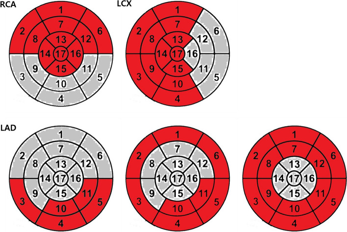
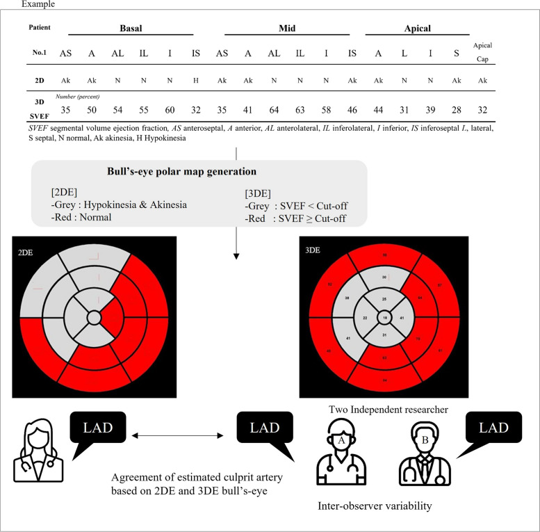
Methods: This retrospective study included 33 patients with ischemic heart disease and single-vessel territory RWMA confirmed on coronary angiography. RWMA was visually assessed using 2DE, generating 17-segment bull's-eye polar maps, and 3DE. In the 3DE study, two independent observers analyzed segmental volumes and segmental volume ejection fractions (SVEFs) using QLAB 3D quantification software. The optimal SVEF cutoff value differentiating normal from abnormal was determined using receiver operating curve analysis. The accuracy of 3DE in predicting culprit coronary arteries was compared with that of 2DE using Cohen κ coefficients, which also were used for interobserver and intraobserver variability assessments.
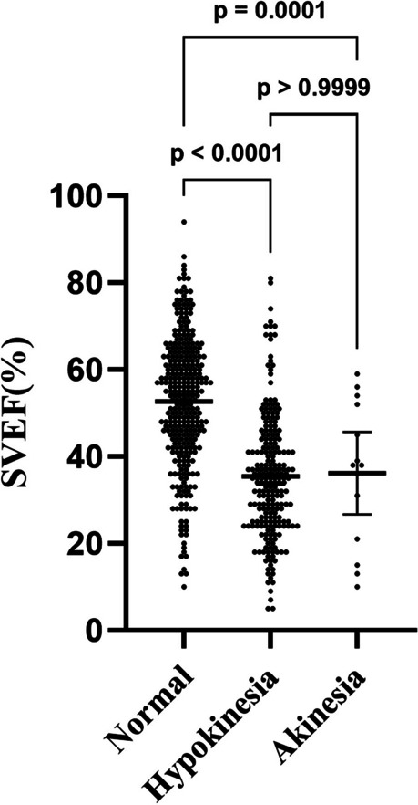
Results: Mean 3DE SVEFs were significantly lower in segments showing RWMA on 2DE. The optimal SVEF cutoff value was 44%, with sensitivity of 75.0% and specificity of 73.9% (area under the curve, 0.801; 95% CI, 0.763-0.838; P < 0.001). The reliability of 3DE-derived bull's-eye predictions of culprit coronary arteries was 81.8% (κ = 0.672; 95% CI, 0.555-0.789; P < 0.001). Interobserver and intraobserver variabilities were 97.0% (κ = 0.947; 95% CI, 0.894-1.00; P < 0.001) and 93.9% (κ = 0.897; 95% CI, 0.827-0.967; P < 0.001), respectively.
Conclusions: The 3DE segmental volume analysis effectively quantified regional left ventricle function and aligned well with 2DE and coronary angiography findings in predicting culprit coronary arteries. Thus, 3DE segmental volume analysis can serve as a quantitative and objective tool for RWMA assessment in patients with ischemic heart disease.

 求助内容:
求助内容: 应助结果提醒方式:


