Molecular architectures of centrosomes in C. elegans embryos visualized by cryo-electron tomography
IF 10.7
1区 生物学
Q1 CELL BIOLOGY
引用次数: 0
Abstract
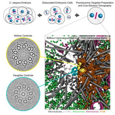
Centrosomes organize microtubules that are essential for mitotic divisions in animal cells. They consist of centrioles surrounded by pericentriolar material (PCM). Questions related to mechanisms of centriole assembly, PCM organization, and spindle microtubule formation remain unanswered, partly due to limited availability of molecular-resolution structural data inside cells. Here, we use cryo-electron tomography to visualize centrosomes across the cell cycle in cells isolated from C. elegans embryos. We describe a pseudo-timeline of centriole assembly and identify distinct structural features in both mother and daughter centrioles. We find that centrioles and PCM microtubules differ in protofilament number (13 versus 11), which could be explained by atypical γ-tubulin ring complexes with 11-fold symmetry identified at the minus ends of short PCM microtubule segments. We further characterize a porous and disordered network that forms the interconnected PCM. Thus, our work builds a three-dimensional structural atlas that helps explain how centrosomes assemble, grow, and achieve function.

用冷冻电子断层扫描观察秀丽隐杆线虫胚胎中心体的分子结构
中心体组织对动物细胞有丝分裂至关重要的微管。它们由中心粒被中心粒周围物质(PCM)包围组成。中心粒组装、PCM组织和纺锤体微管形成机制的相关问题仍未得到解答,部分原因是细胞内分子分辨率结构数据的可用性有限。在这里,我们使用低温电子断层扫描来观察秀丽隐杆线虫胚胎细胞中中心体的细胞周期。我们描述了中心粒组装的伪时间线,并确定了母中心粒和子中心粒的不同结构特征。我们发现中心粒和PCM微管的原丝数不同(13比11),这可能是由于在短PCM微管片段的负端发现了非典型的11倍对称γ-微管蛋白环复合物。我们进一步表征了形成相互连接的PCM的多孔和无序网络。因此,我们的工作建立了一个三维结构图谱,有助于解释中心体如何组装、生长和实现功能。
本文章由计算机程序翻译,如有差异,请以英文原文为准。
求助全文
约1分钟内获得全文
求助全文
来源期刊

Developmental cell
生物-发育生物学
CiteScore
18.90
自引率
1.70%
发文量
203
审稿时长
3-6 weeks
期刊介绍:
Developmental Cell, established in 2001, is a comprehensive journal that explores a wide range of topics in cell and developmental biology. Our publication encompasses work across various disciplines within biology, with a particular emphasis on investigating the intersections between cell biology, developmental biology, and other related fields. Our primary objective is to present research conducted through a cell biological perspective, addressing the essential mechanisms governing cell function, cellular interactions, and responses to the environment. Moreover, we focus on understanding the collective behavior of cells, culminating in the formation of tissues, organs, and whole organisms, while also investigating the consequences of any malfunctions in these intricate processes.
 求助内容:
求助内容: 应助结果提醒方式:


