Devalla Venu Babu, Srinidhi V Ballullaya, Pushpa S, Neha Taufin, Pilli Sai Naveen
{"title":"Nanoparticles Induced Biomimetic Remineralization of Acid-Etched Dentin.","authors":"Devalla Venu Babu, Srinidhi V Ballullaya, Pushpa S, Neha Taufin, Pilli Sai Naveen","doi":"10.30476/dentjods.2024.98928.2117","DOIUrl":null,"url":null,"abstract":"<p><strong>Statement of the problem: </strong>Dentin bonding with etch-and-rinse adhesives involves demineralizing the 5-8µm of the surface dentin to create micro space for resin infiltration. The presence of continuous fluid movement in dentin tubules and positive pulpal pressure prevents complete water replacement by resin monomers. This results in areas of demineralized dentin, which contain collagen fibers without resin infiltration. The exposed collage fibers are subjected to enzymatic degradation leading to less durable hybrid layer.</p><p><strong>Purpose: </strong>The aim of this study was to evaluate the remineralizing effect of the nanoparticles on the resin dentin bonding interface.</p><p><strong>Materials and method: </strong>The three experimental remineralizing nanoparticles were characterized for their morphology, size, and composition. A total of 48 extracted non-carious human third molar teeth were sectioned at 2 mm below the cemento enamel junction. Class I cavity was prepared and the tooth samples were placed in an intra pulpal pressure simulation device. After etching of the prepared cavity, the samples were randomly divided into four groups (n=10) as follows: (1) control group(c) (n=10) (2) Nano-hydroxyapatite (nHAP) (n=10) (3) Chitosan-nanohydroxyapatite (Chi-nHAP) (n=10) (4) Mesoporous silica-hydrox-yapatite (MS-nHAP) (n=10). After 30 days remineralization period, the samples were evaluated for micro tensile bond strength, hybrid layer morphology, and mineral composition of the hybrid layer. The results were analyzed statistically by one-way ANOVA and Tukey's multiple post hoc tests.</p><p><strong>Results: </strong>Scanning electron microscopic observation of nanoparticles revealed irregular particle shapes with calcium phosphate ratio of 1.60. The zeta analyzer showed a mean diameter of 161.0 nm, 323.0nm, 185.0nm for nHAP, Chi-nHAP, and MS-nHAP respectively. Post hoc Bonferroni test revealed significantly higher bond strength for nHAP, Chi-nHAP, and MS-nHAP when compared to control group. MS-nHAP resulted in the uniform deposition of apatite crystal on the surface without any evidence of dentinal tubules openings and had higher mineral to matrix ratio compared to other groups.</p><p><strong>Conclusion: </strong>MS-nHAP nanoparticles can be considered as a reliable source of calcium and phosphate for biomimetic remineralization of hybrid layer. Application of nanoparticle remineralization precursors before application of dentin bonding agents results in remeralization of exposed collagen fibers thereby improving the clinical longevity of hybrid layer.</p>","PeriodicalId":73702,"journal":{"name":"Journal of dentistry (Shiraz, Iran)","volume":"25 4","pages":"359-368"},"PeriodicalIF":0.0000,"publicationDate":"2024-12-01","publicationTypes":"Journal Article","fieldsOfStudy":null,"isOpenAccess":false,"openAccessPdf":"https://www.ncbi.nlm.nih.gov/pmc/articles/PMC11662173/pdf/","citationCount":"0","resultStr":null,"platform":"Semanticscholar","paperid":null,"PeriodicalName":"Journal of dentistry (Shiraz, Iran)","FirstCategoryId":"1085","ListUrlMain":"https://doi.org/10.30476/dentjods.2024.98928.2117","RegionNum":0,"RegionCategory":null,"ArticlePicture":[],"TitleCN":null,"AbstractTextCN":null,"PMCID":null,"EPubDate":"","PubModel":"","JCR":"","JCRName":"","Score":null,"Total":0}
引用次数: 0
Abstract
Statement of the problem: Dentin bonding with etch-and-rinse adhesives involves demineralizing the 5-8µm of the surface dentin to create micro space for resin infiltration. The presence of continuous fluid movement in dentin tubules and positive pulpal pressure prevents complete water replacement by resin monomers. This results in areas of demineralized dentin, which contain collagen fibers without resin infiltration. The exposed collage fibers are subjected to enzymatic degradation leading to less durable hybrid layer.
Purpose: The aim of this study was to evaluate the remineralizing effect of the nanoparticles on the resin dentin bonding interface.
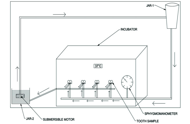
Materials and method: The three experimental remineralizing nanoparticles were characterized for their morphology, size, and composition. A total of 48 extracted non-carious human third molar teeth were sectioned at 2 mm below the cemento enamel junction. Class I cavity was prepared and the tooth samples were placed in an intra pulpal pressure simulation device. After etching of the prepared cavity, the samples were randomly divided into four groups (n=10) as follows: (1) control group(c) (n=10) (2) Nano-hydroxyapatite (nHAP) (n=10) (3) Chitosan-nanohydroxyapatite (Chi-nHAP) (n=10) (4) Mesoporous silica-hydrox-yapatite (MS-nHAP) (n=10). After 30 days remineralization period, the samples were evaluated for micro tensile bond strength, hybrid layer morphology, and mineral composition of the hybrid layer. The results were analyzed statistically by one-way ANOVA and Tukey's multiple post hoc tests.
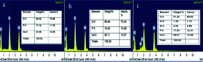
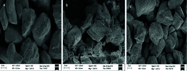
Results: Scanning electron microscopic observation of nanoparticles revealed irregular particle shapes with calcium phosphate ratio of 1.60. The zeta analyzer showed a mean diameter of 161.0 nm, 323.0nm, 185.0nm for nHAP, Chi-nHAP, and MS-nHAP respectively. Post hoc Bonferroni test revealed significantly higher bond strength for nHAP, Chi-nHAP, and MS-nHAP when compared to control group. MS-nHAP resulted in the uniform deposition of apatite crystal on the surface without any evidence of dentinal tubules openings and had higher mineral to matrix ratio compared to other groups.
Conclusion: MS-nHAP nanoparticles can be considered as a reliable source of calcium and phosphate for biomimetic remineralization of hybrid layer. Application of nanoparticle remineralization precursors before application of dentin bonding agents results in remeralization of exposed collagen fibers thereby improving the clinical longevity of hybrid layer.

 求助内容:
求助内容: 应助结果提醒方式:


