RORc-expressing immune cells negatively regulate tertiary lymphoid structure formation and support their pro-tumorigenic functions
IF 33
1区 医学
Q1 GASTROENTEROLOGY & HEPATOLOGY
引用次数: 0
Abstract
Background & Aims
RORc-expressing immune cells play important roles in inflammation, autoimmune disease and cancer. They are required for lymphoid organogenesis and have been implicated in tertiary lymphoid structure (TLS) formation. TLSs are formed in many cancer types and have been correlated with better prognosis and response to immunotherapy. In liver cancer, some TLSs are pro-tumorigenic as they harbor tumor progenitor cells and support their growth. The processes involved in TLS development and acquisition of pro- or anti-tumorigenic roles are largely unknown. This study aims to explore the role of RORc-expressing cells in TLS development in the context of inflammation-associated liver cancer.
Methods
IKKβ(EE)Hep mice, exhibiting chronic liver inflammation, TLS formation and liver cancer, were crossed with RORc knockout mice to explore RORc’s effect on TLS and tumor formation. TLS phenotypes were analyzed using transcriptional, proteomic, and immunohistochemical techniques. CD4, CD8, and B-cell depletions were used to assess their contribution to liver TLS and tumor formation.
Results
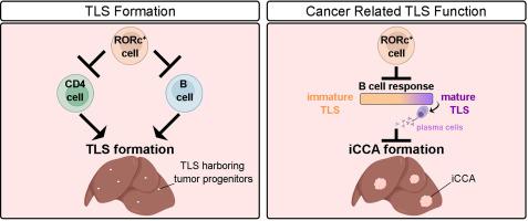
RORc-expressing cells are detected within TLSs of both human patients and mice developing intrahepatic cholangiocarcinoma. In mice, these cells negatively regulate TLS formation, as excess TLSs form in their absence. CD4 cells are essential for liver TLS formation, while B cells are required for TLS formation specifically in the absence of RORc-expressing cells. Importantly, in chronically inflamed livers lacking RORc-expressing cells, TLSs become anti-tumorigenic, reducing tumor load. Anti-tumorigenic TLSs revealed enrichment of exhausted CD8 cells with effector functions, germinal center B cells and plasma cells. B cells are key in limiting tumor development, possibly via tumor-directed antibodies.
Conclusions
RORc-expressing cells negatively regulate B-cell responses and facilitate the pro-tumorigenic functions of hepatic TLSs.
Impact and implications
RORc-expressing immune cells play critical roles in immune regulation, yet their specific influence on tertiary lymphoid structures (TLSs) in liver pathology and cancer has not been elucidated. Our study reveals that RORc-expressing cells act as negative regulators of TLS formation and shape the immune microenvironment in a manner that promotes tumor development. In the absence of RORc-expressing cells, TLSs not only increase in number but also acquire anti-tumorigenic properties. These findings suggest that RORc-expressing cells serve as key modulators of liver immune dynamics, with potential implications for the use of RORc as a biomarker to differentiate between pro- and anti-tumorigenic immune environments and as a target for manipulating TLS abundance and phenotype in liver cancer.


表达RORc的免疫细胞负性调节三级淋巴结构的形成并支持其促肿瘤功能
背景与目的表达rorc的免疫细胞在炎症、自身免疫性疾病和癌症中发挥重要作用。它们是淋巴器官发生所必需的,并与三级淋巴结构(TLS)的形成有关。TLSs在许多癌症类型中形成,并与更好的预后和免疫治疗反应相关。在肝癌中,一些TLSs具有致瘤性,因为它们含有肿瘤祖细胞并支持其生长。涉及TLS发展和获得促或抗肿瘤作用的过程在很大程度上是未知的。方法将表现慢性肝脏炎症、TLS形成和肝癌的sikk β(EE)Hep小鼠与RORc敲除小鼠杂交,探讨RORc对TLS和肿瘤形成的影响。使用转录、蛋白质组学和免疫组织化学技术分析TLS表型。使用CD4、CD8和B细胞消耗来评估它们对肝脏TLS和肿瘤形成的贡献。结果在人类和小鼠肝内胆管癌患者的TLSs中均检测到表达rorc的细胞。在小鼠中,这些细胞负向调节TLS的形成,因为在缺乏它们的情况下会形成过量的TLS。CD4细胞是肝脏TLS形成的必需细胞,而在缺乏rorc表达细胞的情况下,TLS的形成需要B细胞。重要的是,在缺乏rorc表达细胞的慢性炎症肝脏中,TLSs具有抗致瘤性,降低了肿瘤负荷。抗肿瘤TLSs显示具有效应功能的耗尽CD8细胞、生发中心B细胞和浆细胞富集。B细胞是限制肿瘤发展的关键,可能是通过肿瘤定向抗体。结论表达sorc的细胞负向调节B细胞的反应,促进肝脏TLSs的致瘤功能。
本文章由计算机程序翻译,如有差异,请以英文原文为准。
求助全文
约1分钟内获得全文
求助全文
来源期刊

Journal of Hepatology
医学-胃肠肝病学
CiteScore
46.10
自引率
4.30%
发文量
2325
审稿时长
30 days
期刊介绍:
The Journal of Hepatology is the official publication of the European Association for the Study of the Liver (EASL). It is dedicated to presenting clinical and basic research in the field of hepatology through original papers, reviews, case reports, and letters to the Editor. The Journal is published in English and may consider supplements that pass an editorial review.
 求助内容:
求助内容: 应助结果提醒方式:


