Proteolytic Enzyme Activity and mRNA Expressions of Amino Acid and Peptide Transporter Genes in Tissues of Nile tilapia (Oreochromis niloticus) Exposed to Different Salinities
Emmanuel O. Kombat, Godwin Abakari, Abdul-Razak Salifu, Elliot H. Alhassan, Jin-Liang Zhao
{"title":"Proteolytic Enzyme Activity and mRNA Expressions of Amino Acid and Peptide Transporter Genes in Tissues of Nile tilapia (Oreochromis niloticus) Exposed to Different Salinities","authors":"Emmanuel O. Kombat, Godwin Abakari, Abdul-Razak Salifu, Elliot H. Alhassan, Jin-Liang Zhao","doi":"10.1002/aff2.70024","DOIUrl":null,"url":null,"abstract":"<p>This study examined the effects of different salinity levels and exposure time on the activity of proteolytic enzymes and the mRNA expression of amino acid (AA) and peptide transporter genes in various fish tissues of Nile tilapia (<i>Oreochromis niloticus</i>). <i>O. niloticus</i> juveniles, weighing 25.30 ± 4.82 g, were subjected to salt concentrations of 0, 6, 12, 18 and 24 g/L for 30 days. After being exposed to salt for 3 h (acute exposure) and 30 days (chronic exposure), fish tissues (liver, gills and intestines) were sampled. Real-time quantitative PCR (RT-qPCR) was used to determine the mRNA expressions of peptide and AA transporter genes, while the activities of proteolytic enzymes were assessed using enzyme-linked immunosorbent assay (ELISA) test kits. Salinity and exposure time had no significant impact (<i>p</i> > 0.05) on the proteolytic enzyme activity in both intestine and liver tissues of <i>O. niloticus</i>, except for elastase and protease at 24 g/L salinity. PepT1a mRNA gene expression in the fish gut was observed to be considerably elevated (<i>p</i> < 0.05) at both exposure times with increasing salinities. PepT1b did not exhibit any appreciable alterations (<i>p</i> < 0.05) in the gills or intestines at the various salinities. In the intestine, the expression of PepT2 was significantly upregulated (<i>p</i> < 0.05) with rising salinity for chronic exposure but remained unchanged for acute exposure. In contrast, the expression of PepT2 rose substantially (<i>p</i> < 0.05) in the gills with rising salinity for acute exposure and downregulated for chronic exposure. Both exposure time and salinity significantly impacted (<i>p</i> < 0.05) the expression of slc3a1 in the gut. There were significant variations (<i>p</i> < 0.05) in slc3a9 expression in the gut and gill at all salinities for both acute and chronic exposures. In contrast, substantial variations in slc3a19 expression were seen in the intestine at various salinities for acute exposure but not for chronic exposure. The gills displayed significant differences at all salinities for both exposure times.</p>","PeriodicalId":100114,"journal":{"name":"Aquaculture, Fish and Fisheries","volume":"4 6","pages":""},"PeriodicalIF":1.9000,"publicationDate":"2024-12-09","publicationTypes":"Journal Article","fieldsOfStudy":null,"isOpenAccess":false,"openAccessPdf":"https://onlinelibrary.wiley.com/doi/epdf/10.1002/aff2.70024","citationCount":"0","resultStr":null,"platform":"Semanticscholar","paperid":null,"PeriodicalName":"Aquaculture, Fish and Fisheries","FirstCategoryId":"1085","ListUrlMain":"https://onlinelibrary.wiley.com/doi/10.1002/aff2.70024","RegionNum":0,"RegionCategory":null,"ArticlePicture":[],"TitleCN":null,"AbstractTextCN":null,"PMCID":null,"EPubDate":"","PubModel":"","JCR":"Q3","JCRName":"FISHERIES","Score":null,"Total":0}
引用次数: 0
Abstract
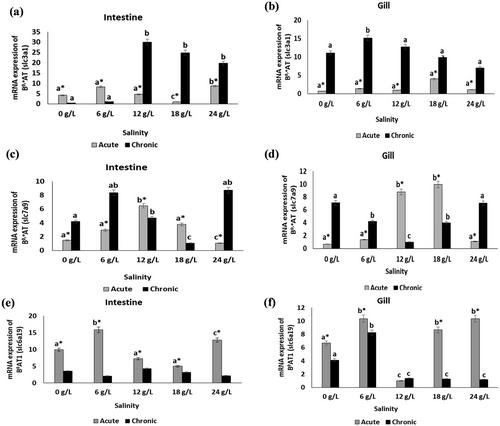
This study examined the effects of different salinity levels and exposure time on the activity of proteolytic enzymes and the mRNA expression of amino acid (AA) and peptide transporter genes in various fish tissues of Nile tilapia (Oreochromis niloticus). O. niloticus juveniles, weighing 25.30 ± 4.82 g, were subjected to salt concentrations of 0, 6, 12, 18 and 24 g/L for 30 days. After being exposed to salt for 3 h (acute exposure) and 30 days (chronic exposure), fish tissues (liver, gills and intestines) were sampled. Real-time quantitative PCR (RT-qPCR) was used to determine the mRNA expressions of peptide and AA transporter genes, while the activities of proteolytic enzymes were assessed using enzyme-linked immunosorbent assay (ELISA) test kits. Salinity and exposure time had no significant impact (p > 0.05) on the proteolytic enzyme activity in both intestine and liver tissues of O. niloticus, except for elastase and protease at 24 g/L salinity. PepT1a mRNA gene expression in the fish gut was observed to be considerably elevated (p < 0.05) at both exposure times with increasing salinities. PepT1b did not exhibit any appreciable alterations (p < 0.05) in the gills or intestines at the various salinities. In the intestine, the expression of PepT2 was significantly upregulated (p < 0.05) with rising salinity for chronic exposure but remained unchanged for acute exposure. In contrast, the expression of PepT2 rose substantially (p < 0.05) in the gills with rising salinity for acute exposure and downregulated for chronic exposure. Both exposure time and salinity significantly impacted (p < 0.05) the expression of slc3a1 in the gut. There were significant variations (p < 0.05) in slc3a9 expression in the gut and gill at all salinities for both acute and chronic exposures. In contrast, substantial variations in slc3a19 expression were seen in the intestine at various salinities for acute exposure but not for chronic exposure. The gills displayed significant differences at all salinities for both exposure times.

 求助内容:
求助内容: 应助结果提醒方式:


