Christy Buckley, Caroline V Fulkerson, Maxime Derre, Andrew Woolcock, Masahiro Murakami
{"title":"Hepatic parenchymal hypoattenuation in dogs with diabetes mellitus on computed tomography consistent with hepatic steatosis.","authors":"Christy Buckley, Caroline V Fulkerson, Maxime Derre, Andrew Woolcock, Masahiro Murakami","doi":"10.1111/vru.13464","DOIUrl":null,"url":null,"abstract":"<p><p>Hypoattenuation of the liver, consistent with hepatic steatosis or lipidosis, has been reported in veterinary patients. In people, measuring CT hepatic attenuation is diagnostic for hepatic steatosis, and hypoattenuation of the liver is defined as absolute if less than 40 HU or relative if the liver is 10 HU less than the spleen. The purpose of this study is to describe hepatic parenchymal attenuation in dogs with diabetes mellitus with or without diabetic ketosis (DK) or diabetic ketoacidosis (DKA), using the above categorization for absolute and relative hypoattenuation, as with humans. We hypothesized dogs with DK or DKA were more likely to have hypoattenuating livers. Twenty-seven diabetic dogs were included; fifteen were categorized in Group 1 as without DK or DKA, six in Group 2 as DK, and six in Group 3 as DKA. In Group 3, four of six dogs had absolute and relative hypoattenuating livers. Three of these were visually hypoattenuating to the vasculature, with one having negative attenuation and a histopathologic diagnosis of severe hepatic lipidosis. In Group 2, four of six dogs had relative hypoattenuating livers. In Group 1, only one of 15 dogs had a relatively hypoattenuating liver. Groups 2 and 3 had significantly lower absolute liver attenuation than Group 1. Presumed hepatic steatosis was present on CT and was more common with DK or DKA. These findings may help provide hepatic sampling recommendations and alter patient prognosis. Further research is needed to establish absolute and relative liver attenuation in dogs with correlation to histopathology and patient outcome.</p>","PeriodicalId":23581,"journal":{"name":"Veterinary Radiology & Ultrasound","volume":"66 1","pages":"e13464"},"PeriodicalIF":1.5000,"publicationDate":"2025-01-01","publicationTypes":"Journal Article","fieldsOfStudy":null,"isOpenAccess":false,"openAccessPdf":"https://www.ncbi.nlm.nih.gov/pmc/articles/PMC11649884/pdf/","citationCount":"0","resultStr":null,"platform":"Semanticscholar","paperid":null,"PeriodicalName":"Veterinary Radiology & Ultrasound","FirstCategoryId":"97","ListUrlMain":"https://doi.org/10.1111/vru.13464","RegionNum":2,"RegionCategory":"农林科学","ArticlePicture":[],"TitleCN":null,"AbstractTextCN":null,"PMCID":null,"EPubDate":"","PubModel":"","JCR":"Q2","JCRName":"VETERINARY SCIENCES","Score":null,"Total":0}
引用次数: 0
Abstract
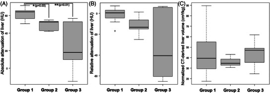
Hypoattenuation of the liver, consistent with hepatic steatosis or lipidosis, has been reported in veterinary patients. In people, measuring CT hepatic attenuation is diagnostic for hepatic steatosis, and hypoattenuation of the liver is defined as absolute if less than 40 HU or relative if the liver is 10 HU less than the spleen. The purpose of this study is to describe hepatic parenchymal attenuation in dogs with diabetes mellitus with or without diabetic ketosis (DK) or diabetic ketoacidosis (DKA), using the above categorization for absolute and relative hypoattenuation, as with humans. We hypothesized dogs with DK or DKA were more likely to have hypoattenuating livers. Twenty-seven diabetic dogs were included; fifteen were categorized in Group 1 as without DK or DKA, six in Group 2 as DK, and six in Group 3 as DKA. In Group 3, four of six dogs had absolute and relative hypoattenuating livers. Three of these were visually hypoattenuating to the vasculature, with one having negative attenuation and a histopathologic diagnosis of severe hepatic lipidosis. In Group 2, four of six dogs had relative hypoattenuating livers. In Group 1, only one of 15 dogs had a relatively hypoattenuating liver. Groups 2 and 3 had significantly lower absolute liver attenuation than Group 1. Presumed hepatic steatosis was present on CT and was more common with DK or DKA. These findings may help provide hepatic sampling recommendations and alter patient prognosis. Further research is needed to establish absolute and relative liver attenuation in dogs with correlation to histopathology and patient outcome.
期刊介绍:
Veterinary Radiology & Ultrasound is a bimonthly, international, peer-reviewed, research journal devoted to the fields of veterinary diagnostic imaging and radiation oncology. Established in 1958, it is owned by the American College of Veterinary Radiology and is also the official journal for six affiliate veterinary organizations. Veterinary Radiology & Ultrasound is represented on the International Committee of Medical Journal Editors, World Association of Medical Editors, and Committee on Publication Ethics.
The mission of Veterinary Radiology & Ultrasound is to serve as a leading resource for high quality articles that advance scientific knowledge and standards of clinical practice in the areas of veterinary diagnostic radiology, computed tomography, magnetic resonance imaging, ultrasonography, nuclear imaging, radiation oncology, and interventional radiology. Manuscript types include original investigations, imaging diagnosis reports, review articles, editorials and letters to the Editor. Acceptance criteria include originality, significance, quality, reader interest, composition and adherence to author guidelines.

 求助内容:
求助内容: 应助结果提醒方式:


