In Vivo Surface-Enhanced Transmission Raman Spectroscopy and Impact of Frozen Biological Tissues on Lesion Depth Prediction
IF 16
1区 材料科学
Q1 CHEMISTRY, MULTIDISCIPLINARY
引用次数: 0
Abstract
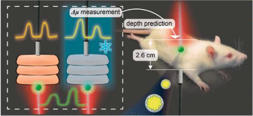
Plasmonic surface-enhanced transmission Raman spectroscopy (SETRS) has emerged as a promising optical technique for detecting and predicting the depths of deep-seated lesions in biological tissues. However, in vivo studies using SETRS are scarce and typically show shallow penetration depths. Moreover, the optical parameters used in the prediction process are often derived from frozen samples and there is limited understanding of how freezing affects the optical properties of biological tissues and the accuracy of depth prediction in living models. In this work, we conduct in vivo SETRS measurements on thick abdominal tissue region of the live rats to investigate the impact of freezing on the measured optical properties for the purpose of depth prediction. First, we fabricated ultrahigh bright surface-enhanced Raman spectroscopy (SERS) nanotags and utilized a custom transmission Raman system. We then measured the change of optical attenuation at two different wavelengths (Δμ) for four types of rat tissues (including skin, fat, muscle, and liver) following freezing. The freezing process dramatically affects Δμ values, even after only 1 day of freezing. In contrast, Δμ values obtained from fresh samples enable precise localization of SERS lesion phantoms in the live rat with only 5% deviation. The total thickness of the live rat is 2.6 cm, which, to the best of our knowledge, is the highest value of in vivo SETRS studies so far. This work helps to fill the gap in the SERS field of tissue localization and optical coefficient studies in highly heterogeneous tissues, and demonstrates the potential of the SETRS technique to achieve precise clinical localization of deep lesions.

体内表面增强透射拉曼光谱和冷冻生物组织对病变深度预测的影响
质子表面增强透射拉曼光谱(SETRS)已成为一种很有前途的光学技术,可用于检测和预测生物组织深层病变的深度。然而,使用 SETRS 进行的活体研究很少,通常显示的穿透深度较浅。此外,预测过程中使用的光学参数通常来自冷冻样本,人们对冷冻如何影响生物组织的光学特性以及活体模型深度预测的准确性了解有限。在这项工作中,我们对活体大鼠腹部厚组织区域进行了活体 SETRS 测量,以研究冷冻对所测光学特性的影响,从而达到深度预测的目的。首先,我们制作了超高亮度表面增强拉曼光谱(SERS)纳米标签,并利用定制的透射拉曼系统。然后,我们测量了四种大鼠组织(包括皮肤、脂肪、肌肉和肝脏)在冷冻后两种不同波长(Δμ)的光衰减变化。冷冻过程会显著影响Δμ值,即使冷冻仅 1 天后也是如此。相比之下,从新鲜样本中获得的Δμ值能精确定位活体大鼠的 SERS 病变模型,偏差仅为 5%。活体大鼠的总厚度为 2.6 厘米,据我们所知,这是迄今为止活体 SETRS 研究的最高值。这项工作有助于填补 SERS 领域在高度异质组织中进行组织定位和光学系数研究的空白,并展示了 SETRS 技术在实现深部病变精确临床定位方面的潜力。
本文章由计算机程序翻译,如有差异,请以英文原文为准。
求助全文
约1分钟内获得全文
求助全文
来源期刊

ACS Nano
工程技术-材料科学:综合
CiteScore
26.00
自引率
4.10%
发文量
1627
审稿时长
1.7 months
期刊介绍:
ACS Nano, published monthly, serves as an international forum for comprehensive articles on nanoscience and nanotechnology research at the intersections of chemistry, biology, materials science, physics, and engineering. The journal fosters communication among scientists in these communities, facilitating collaboration, new research opportunities, and advancements through discoveries. ACS Nano covers synthesis, assembly, characterization, theory, and simulation of nanostructures, nanobiotechnology, nanofabrication, methods and tools for nanoscience and nanotechnology, and self- and directed-assembly. Alongside original research articles, it offers thorough reviews, perspectives on cutting-edge research, and discussions envisioning the future of nanoscience and nanotechnology.
文献相关原料
公司名称
产品信息
阿拉丁
Cetyltrimethylammonium chloride (CTAC)
阿拉丁
ascorbic acid (AA)
阿拉丁
silver nitrate (AgNO3)
阿拉丁
agarose
 求助内容:
求助内容: 应助结果提醒方式:


