{"title":"The value of dual-energy computed tomography angiography-based virtual monoenergetic imaging for evaluations after cerebral aneurysm clipping.","authors":"Zhihua Lu, Suying Wu, Feijian Wu, Qingdong Jin, Qingjing Huang, Baoteng Zhang","doi":"10.4274/dir.2024.242975","DOIUrl":null,"url":null,"abstract":"<p><strong>Purpose: </strong>This study aimed to research the optimal energy range of dual-energy computed tomography angiography (DECTA)-based virtual monoenergetic imaging (VMI) for evaluations after cerebral aneurysm clipping.</p><p><strong>Methods: </strong>Sixty patients who underwent DECTA after cerebral aneurysm clipping were analyzed retrospectively. Conventional computed tomography angiography (CTA) was compared with VMIs at 60, 70, 80, 90, and 100 keV. The mean attenuation and standard deviation values within the regions of interest placed in the brain parenchyma and arteries with the worst artifact were measured, respectively. The ΔCT and artifact index (AI) values were calculated to assess the artifact severity. The contrast-to-noise ratio (CNR) was calculated to assess vascular contrast. Two radiologists assessed brain parenchyma and cerebrovascular scores qualitatively using a five-point Likert scale.</p><p><strong>Results: </strong>Quantitative analysis showed that the artifacts of VMIs were significantly reduced compared with conventional CTA (<i>P</i> ≤ 0.014), except for the ΔCT and AI of 60 keV and the ΔCT of 70 keV. However, there was no significant difference in the vascular contrast on VMIs compared with conventional CTA, except for the CNR of 60 keV (<i>P</i> = 0.008). In qualitative analysis, the proportions of brain parenchyma scores and cerebrovascular scores ≥4 on the VMIs of 70 and 80 keV were higher than those of conventional CTA and other VMIs.</p><p><strong>Conclusion: </strong>For the patients who underwent DECTA after cerebral aneurysm clipping, the 70-80 keV VMIs are expected to be the optimal energy range for balancing clip artifacts and visibility of adjacent vessels.</p><p><strong>Clinical significance: </strong>Studying the optimal energy range of DECTA-based VMI for post-operative assessment of aneurysm clipping can reduce metal artifacts in images and increase vascular contrast. This facilitates the follow-up of patients after aneurysm clipping, offers timely and accurate detection of postoperative complications, provides assistance to clinicians in diagnosis and treatment, and improves patient prognosis.</p>","PeriodicalId":11341,"journal":{"name":"Diagnostic and interventional radiology","volume":" ","pages":"264-273"},"PeriodicalIF":1.7000,"publicationDate":"2025-04-28","publicationTypes":"Journal Article","fieldsOfStudy":null,"isOpenAccess":false,"openAccessPdf":"https://www.ncbi.nlm.nih.gov/pmc/articles/PMC12057534/pdf/","citationCount":"0","resultStr":null,"platform":"Semanticscholar","paperid":null,"PeriodicalName":"Diagnostic and interventional radiology","FirstCategoryId":"3","ListUrlMain":"https://doi.org/10.4274/dir.2024.242975","RegionNum":4,"RegionCategory":"医学","ArticlePicture":[],"TitleCN":null,"AbstractTextCN":null,"PMCID":null,"EPubDate":"2024/12/16 0:00:00","PubModel":"Epub","JCR":"Q3","JCRName":"RADIOLOGY, NUCLEAR MEDICINE & MEDICAL IMAGING","Score":null,"Total":0}
引用次数: 0
Abstract
Purpose: This study aimed to research the optimal energy range of dual-energy computed tomography angiography (DECTA)-based virtual monoenergetic imaging (VMI) for evaluations after cerebral aneurysm clipping.
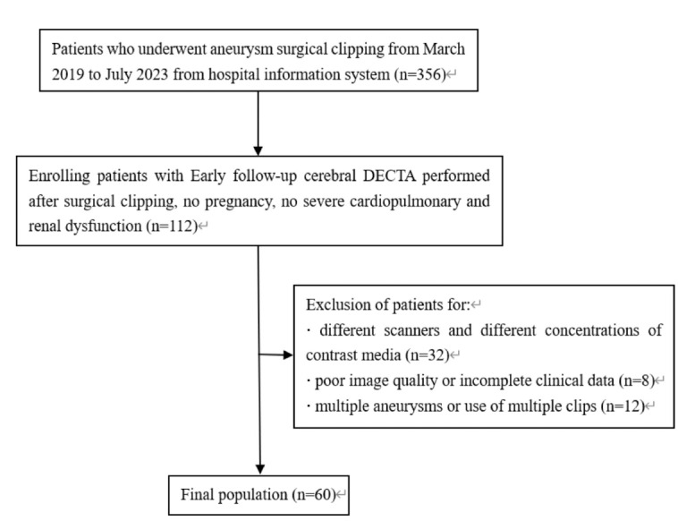
Methods: Sixty patients who underwent DECTA after cerebral aneurysm clipping were analyzed retrospectively. Conventional computed tomography angiography (CTA) was compared with VMIs at 60, 70, 80, 90, and 100 keV. The mean attenuation and standard deviation values within the regions of interest placed in the brain parenchyma and arteries with the worst artifact were measured, respectively. The ΔCT and artifact index (AI) values were calculated to assess the artifact severity. The contrast-to-noise ratio (CNR) was calculated to assess vascular contrast. Two radiologists assessed brain parenchyma and cerebrovascular scores qualitatively using a five-point Likert scale.
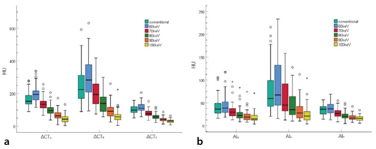
Results: Quantitative analysis showed that the artifacts of VMIs were significantly reduced compared with conventional CTA (P ≤ 0.014), except for the ΔCT and AI of 60 keV and the ΔCT of 70 keV. However, there was no significant difference in the vascular contrast on VMIs compared with conventional CTA, except for the CNR of 60 keV (P = 0.008). In qualitative analysis, the proportions of brain parenchyma scores and cerebrovascular scores ≥4 on the VMIs of 70 and 80 keV were higher than those of conventional CTA and other VMIs.
Conclusion: For the patients who underwent DECTA after cerebral aneurysm clipping, the 70-80 keV VMIs are expected to be the optimal energy range for balancing clip artifacts and visibility of adjacent vessels.
Clinical significance: Studying the optimal energy range of DECTA-based VMI for post-operative assessment of aneurysm clipping can reduce metal artifacts in images and increase vascular contrast. This facilitates the follow-up of patients after aneurysm clipping, offers timely and accurate detection of postoperative complications, provides assistance to clinicians in diagnosis and treatment, and improves patient prognosis.
期刊介绍:
Diagnostic and Interventional Radiology (Diagn Interv Radiol) is the open access, online-only official publication of Turkish Society of Radiology. It is published bimonthly and the journal’s publication language is English.
The journal is a medium for original articles, reviews, pictorial essays, technical notes related to all fields of diagnostic and interventional radiology.

 求助内容:
求助内容: 应助结果提醒方式:


