Fan Huo, Huixia Liu, Weiqi Guo, Hanye Kang, Huihui Zhang, Roman Jashenko, Rong Ji, Hongxia Hu
求助PDF
{"title":"Proliferation dynamic of Paranosema locustae after infection and histopathogenic features on Locusta migratoria","authors":"Fan Huo, Huixia Liu, Weiqi Guo, Hanye Kang, Huihui Zhang, Roman Jashenko, Rong Ji, Hongxia Hu","doi":"10.1002/ps.8601","DOIUrl":null,"url":null,"abstract":"<p><i>Paranosema locustae</i> are specialized parasites of Orthoptera that have been applied widely in the control of grasshoppers in many parts of the world. However, it is slow to kill insects, and how it works in the host is unclear. This work aimed to examine the proliferation of <i>P. locustae</i> within locust tissues and characterize the histological alterations occurring in the midgut, hindgut, and gonads of infected <i>Locusta migratoria</i>. The results showed that during the later stage of infection, the reproduction of <i>P. locustae</i> was most prominent in the fat body and salivary glands (10<sup>9.26</sup> and 10<sup>8.91</sup> copies /ug DNA, respectively). In contrast, the load of <i>P. locustae</i> was least in the craw and midgut (10<sup>7.37</sup> and 10<sup>7.58</sup> copies /ug DNA, respectively), illustrating that the proliferation of <i>P. locustae</i> in the body of locusts had a tissue tendency. The histopathological study revealed that lesions in the hindgut occurred prior to those in the midgut, indicating that <i>P. locustae</i> may have a mechanism for survival that enables it to avoid immune responses in specific organs. The testis exhibited earlier lesions compared to the ovaries, and in the advanced stages of infection, the testis harbored a higher load of <i>P. locustae</i> than the ovaries, suggesting a more pronounced impact on the male reproductive organs in comparison to the female ones. The results of our study enhance our comprehension of the rapid growth and disease-causing mechanism of <i>P. locustae</i>, which can serve as a basis for enhancing its ability to kill insects. © 2024 Society of Chemical Industry.</p>","PeriodicalId":218,"journal":{"name":"Pest Management Science","volume":"81 4","pages":"2051-2060"},"PeriodicalIF":3.8000,"publicationDate":"2024-12-16","publicationTypes":"Journal Article","fieldsOfStudy":null,"isOpenAccess":false,"openAccessPdf":"","citationCount":"0","resultStr":null,"platform":"Semanticscholar","paperid":null,"PeriodicalName":"Pest Management Science","FirstCategoryId":"97","ListUrlMain":"https://scijournals.onlinelibrary.wiley.com/doi/10.1002/ps.8601","RegionNum":1,"RegionCategory":"农林科学","ArticlePicture":[],"TitleCN":null,"AbstractTextCN":null,"PMCID":null,"EPubDate":"","PubModel":"","JCR":"Q1","JCRName":"AGRONOMY","Score":null,"Total":0}
引用次数: 0
引用
批量引用
Abstract
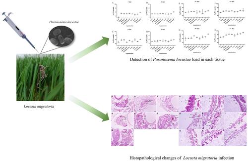
Paranosema locustae are specialized parasites of Orthoptera that have been applied widely in the control of grasshoppers in many parts of the world. However, it is slow to kill insects, and how it works in the host is unclear. This work aimed to examine the proliferation of P. locustae within locust tissues and characterize the histological alterations occurring in the midgut, hindgut, and gonads of infected Locusta migratoria . The results showed that during the later stage of infection, the reproduction of P. locustae was most prominent in the fat body and salivary glands (109.26 and 108.91 copies /ug DNA, respectively). In contrast, the load of P. locustae was least in the craw and midgut (107.37 and 107.58 copies /ug DNA, respectively), illustrating that the proliferation of P. locustae in the body of locusts had a tissue tendency. The histopathological study revealed that lesions in the hindgut occurred prior to those in the midgut, indicating that P. locustae may have a mechanism for survival that enables it to avoid immune responses in specific organs. The testis exhibited earlier lesions compared to the ovaries, and in the advanced stages of infection, the testis harbored a higher load of P. locustae than the ovaries, suggesting a more pronounced impact on the male reproductive organs in comparison to the female ones. The results of our study enhance our comprehension of the rapid growth and disease-causing mechanism of P. locustae , which can serve as a basis for enhancing its ability to kill insects. © 2024 Society of Chemical Industry.


 求助内容:
求助内容: 应助结果提醒方式:


