The Use of the Facial Sinus Wall as Bone Shell Onlay Graft for Maxillary Posterior Ridge Reconstruction: A Retrospective Case Series.

IF 4.8 1区 医学 Q1 DENTISTRY, ORAL SURGERY & MEDICINE
Stefan Krennmair, Michael Weinländer, Michael Malek, Thomas Forstner, Michael Stimmelmayr, Gerald Krennmair
{"title":"The Use of the Facial Sinus Wall as Bone Shell Onlay Graft for Maxillary Posterior Ridge Reconstruction: A Retrospective Case Series.","authors":"Stefan Krennmair, Michael Weinländer, Michael Malek, Thomas Forstner, Michael Stimmelmayr, Gerald Krennmair","doi":"10.1111/clr.14387","DOIUrl":null,"url":null,"abstract":"<p><strong>Purpose: </strong>To evaluate the performance and clinical outcome of vertical and horizontal bone augmentation (VHBA) in posterior maxillary regions combining lateral window sinus floor elevation (LWSFE) with a horizontal bone shell technique applying the maxillary facial sinus wall as a bone plate.</p><p><strong>Materials and methods: </strong>In 18 patients, LWSFE was combined with a horizontal bone shield augmentation procedure utilizing the maxillary facial sinus bone wall as a lateral bone plate. Both the sinus cavity and the lateral bone box created were grafted with a mixture of autogenous bone/venous blood and bovine bone mineral. The primary aim was to assess the performance of combined techniques enabling subsequent implant placement. Using radiographic measurements (preoperative, after VHBA, at implant placement, and at follow-up), bone gain/reduction of augmented horizontal ridge width (HRW) and vertical bone height (VBH) were evaluated. Additionally, clinical outcome assessing implant survival/success rate, marginal bone loss (MBL), and implant health (mucositis/peri-implantitis) was evaluated.</p><p><strong>Results: </strong>For the combined VHBA techniques, HRW and VBH increased significantly (p < 0.001) from preoperative 3.5 ± 1.4 mm/3.6 ± 2.1 mm to 9.7 ± 1.9 mm/18.0 ± 1.6 mm post-augmentation. However, HRW and VBH dimensions decreased up to 8.9 ± 1.8 mm/17.1 ± 1.4 mm at implant placement and 8.6 ± 1.7 mm/16.7 ± 1.3 mm at follow-up evaluation (3.8 ± 1.8 years; p < 0.001, respectively). Augmented bone reduction was significantly higher (-7.7%) between the augmentation procedure and implant placement than in the post-implant-placement period (-2.5%). All implants survived (100%) representing peri-implant MBL of -0.9 ± 0.7 mm, pocket depth of 3.4 + 1.8 mm, and prevalences of 5%/0% for peri-implant mucositis/peri-implantitis.</p><p><strong>Conclusion: </strong>The combination of horizontal bone augmentation using local bone shield transfer from the maxillary facial sinus wall with LWSFE enables sufficient reconstruction of maxillary posterior ridge.</p>","PeriodicalId":10455,"journal":{"name":"Clinical Oral Implants Research","volume":" ","pages":""},"PeriodicalIF":4.8000,"publicationDate":"2024-12-08","publicationTypes":"Journal Article","fieldsOfStudy":null,"isOpenAccess":false,"openAccessPdf":"","citationCount":"0","resultStr":null,"platform":"Semanticscholar","paperid":null,"PeriodicalName":"Clinical Oral Implants Research","FirstCategoryId":"5","ListUrlMain":"https://doi.org/10.1111/clr.14387","RegionNum":1,"RegionCategory":"医学","ArticlePicture":[],"TitleCN":null,"AbstractTextCN":null,"PMCID":null,"EPubDate":"","PubModel":"","JCR":"Q1","JCRName":"DENTISTRY, ORAL SURGERY & MEDICINE","Score":null,"Total":0}
引用次数: 0

Abstract

Purpose: To evaluate the performance and clinical outcome of vertical and horizontal bone augmentation (VHBA) in posterior maxillary regions combining lateral window sinus floor elevation (LWSFE) with a horizontal bone shell technique applying the maxillary facial sinus wall as a bone plate.

Materials and methods: In 18 patients, LWSFE was combined with a horizontal bone shield augmentation procedure utilizing the maxillary facial sinus bone wall as a lateral bone plate. Both the sinus cavity and the lateral bone box created were grafted with a mixture of autogenous bone/venous blood and bovine bone mineral. The primary aim was to assess the performance of combined techniques enabling subsequent implant placement. Using radiographic measurements (preoperative, after VHBA, at implant placement, and at follow-up), bone gain/reduction of augmented horizontal ridge width (HRW) and vertical bone height (VBH) were evaluated. Additionally, clinical outcome assessing implant survival/success rate, marginal bone loss (MBL), and implant health (mucositis/peri-implantitis) was evaluated.

Results: For the combined VHBA techniques, HRW and VBH increased significantly (p < 0.001) from preoperative 3.5 ± 1.4 mm/3.6 ± 2.1 mm to 9.7 ± 1.9 mm/18.0 ± 1.6 mm post-augmentation. However, HRW and VBH dimensions decreased up to 8.9 ± 1.8 mm/17.1 ± 1.4 mm at implant placement and 8.6 ± 1.7 mm/16.7 ± 1.3 mm at follow-up evaluation (3.8 ± 1.8 years; p < 0.001, respectively). Augmented bone reduction was significantly higher (-7.7%) between the augmentation procedure and implant placement than in the post-implant-placement period (-2.5%). All implants survived (100%) representing peri-implant MBL of -0.9 ± 0.7 mm, pocket depth of 3.4 + 1.8 mm, and prevalences of 5%/0% for peri-implant mucositis/peri-implantitis.

Conclusion: The combination of horizontal bone augmentation using local bone shield transfer from the maxillary facial sinus wall with LWSFE enables sufficient reconstruction of maxillary posterior ridge.
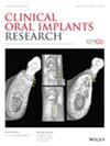
应用面窦壁作为骨壳嵌片重建上颌后嵴:回顾性病例系列。
目的:评价以上颌面窦壁为骨板,结合侧窗窦底提升(LWSFE)和水平骨壳技术在上颌后区进行垂直和水平骨增强(VHBA)的性能和临床效果。材料和方法:18例患者采用LWSFE联合水平骨盾增强术,利用上颌面窦骨壁作为侧骨板。用自体骨/静脉血和牛骨矿物质混合剂移植窦腔和侧骨盒。主要目的是评估联合技术的性能,以便后续植入。通过x线测量(术前、VHBA后、种植体放置时和随访时),评估增强水平脊宽(HRW)和垂直骨高(VBH)的骨增重/减少。此外,评估种植体存活/成功率、边缘骨质流失(MBL)和种植体健康(粘膜炎/种植体周围炎)的临床结果。结果:结合VHBA技术,HRW和VBH明显增加(p)。结论:上颌面窦壁局部骨盾转移水平骨增强术与LWSFE结合,可以充分重建上颌后脊。
本文章由计算机程序翻译,如有差异,请以英文原文为准。
求助全文
约1分钟内获得全文 求助全文
来源期刊
Clinical Oral Implants Research
Clinical Oral Implants Research 医学-工程:生物医学
CiteScore
7.70
自引率
11.60%
发文量
149
审稿时长
3 months
期刊介绍: Clinical Oral Implants Research conveys scientific progress in the field of implant dentistry and its related areas to clinicians, teachers and researchers concerned with the application of this information for the benefit of patients in need of oral implants. The journal addresses itself to clinicians, general practitioners, periodontists, oral and maxillofacial surgeons and prosthodontists, as well as to teachers, academicians and scholars involved in the education of professionals and in the scientific promotion of the field of implant dentistry.
×
引用
GB/T 7714-2015
复制
MLA
复制
APA
复制
导出至
BibTeX EndNote RefMan NoteFirst NoteExpress
×
提示
您的信息不完整,为了账户安全,请先补充。
现在去补充
×
提示
您因"违规操作"
具体请查看互助需知
我知道了
×
提示
确定
请完成安全验证×
copy
已复制链接
快去分享给好友吧!
我知道了
右上角分享
点击右上角分享
0
联系我们:info@booksci.cn Book学术提供免费学术资源搜索服务,方便国内外学者检索中英文文献。致力于提供最便捷和优质的服务体验。 Copyright © 2023 布克学术 All rights reserved.
京ICP备2023020795号-1
ghs 京公网安备 11010802042870号
Book学术文献互助
Book学术文献互助群
群 号:481959085
Book学术官方微信