Hematopoietic Stem Cell Transplantation in Patients With Myelofibrosis and Splanchnic Vein Thrombosis: A Case Series

IF 10.1 1区 医学 Q1 HEMATOLOGY
Portia Smallbone, Mallika Sekhar, Samer A. Srour, Jeremy L. Ramdial, Crystal L. Carmicheal Kusy, Elizabeth J. Shpall, Uday R. Popat
{"title":"Hematopoietic Stem Cell Transplantation in Patients With Myelofibrosis and Splanchnic Vein Thrombosis: A Case Series","authors":"Portia Smallbone, Mallika Sekhar, Samer A. Srour, Jeremy L. Ramdial, Crystal L. Carmicheal Kusy, Elizabeth J. Shpall, Uday R. Popat","doi":"10.1002/ajh.27542","DOIUrl":null,"url":null,"abstract":"<p>Myeloproliferative neoplasms (MPN) are associated with increased risk of splanchnic vein thrombosis (SVT), which contributes to morbidity and mortality. SVT, including portal, superior mesenteric, splenic, and hepatic vein thrombosis, disrupts portal pressures, leading to the development of portal hypertension (PHT) and complications such as varices, splenomegaly and liver failure, which increase the morbidity and mortality. The impact of prior SVT on outcomes of patients undergoing allogeneic hematopoietic stem cell transplantation (HCT) for myelofibrosis (MF) is not well understood. Although a small case series found a strong correlation between SVT and hyperbilirubinemia [<span>1</span>], the data is limited, making the optimal management of anticoagulation, hepatic comorbidities, and PHT sequelae challenging in this patient population. This study evaluates the impact of pre-existing SVT and its treatment on transplant outcomes.</p>\n<p>We screened 334 consecutive patients who underwent HCT for MF at The University of Texas MD Anderson Cancer Center between January 2000 and August 2023 and identified 12 with pre-existing SVT. Patient characteristics, liver and thrombosis-related data, transplant details and outcomes were collected through retrospective review. Model for End-Stage Liver Disease (MELD) scores were calculated using creatinine, bilirubin, INR, and sodium levels to stratify the degree of chronic liver dysfunction. Continuous variables are reported as median and range, while categorical variables as number and percentage. Overall survival was estimated using the Kaplan–Meier method.</p>\n<p>With regards to patient characteristics, the median age was 53 years (range: 39–73). Five patients had primary MF, four had post-essential thrombocythemia (PET-MF), and three had post-polycythaemia vera (PPV-MF). The majority (<i>n</i> = 8, 66.6%) were JAK2 V617F-positive. One patient was MPL positive. Patients received conditioning regimens based on institutional protocols, considering age and comorbidities. Individual characteristics and outcomes for all patients are summarized in Table 1. Other baseline characteristics are seen in Table S1.</p>\n<div>\n<header><span>TABLE 1. </span>Detailed patient characteristics.</header>\n<div tabindex=\"0\">\n<table>\n<thead>\n<tr>\n<th>Patient</th>\n<th>Age at transplant</th>\n<th>Sex</th>\n<th>JAK2 status</th>\n<th>Additional mutations via NGS</th>\n<th>Thrombosis type</th>\n<th>Days SVT prior to transplant</th>\n<th>Splenectomy prior to thrombosis</th>\n<th>Yerdel classification</th>\n<th>Anticoagulation</th>\n<th>Date of transplant</th>\n<th>Conditioning regimen</th>\n</tr>\n</thead>\n<tbody>\n<tr>\n<td>1</td>\n<td>40</td>\n<td>F</td>\n<td>Positive</td>\n<td>No</td>\n<td>Splenic vein thrombosis</td>\n<td>109</td>\n<td>No</td>\n<td>NE</td>\n<td>No</td>\n<td>3-Jul-23</td>\n<td>Bu/Cy</td>\n</tr>\n<tr>\n<td>2</td>\n<td>70</td>\n<td>F</td>\n<td>Positive</td>\n<td>No</td>\n<td>Portal vein, SMV and splenic vein thrombosis</td>\n<td>145</td>\n<td>Yes</td>\n<td>3</td>\n<td>Yes, 120 days</td>\n<td>14-Sep-22</td>\n<td>Flu/Mel</td>\n</tr>\n<tr>\n<td>3</td>\n<td>72</td>\n<td>M</td>\n<td>Positive</td>\n<td>ASXL1, EZH2, GATA2, SF3B1</td>\n<td>Portal vein thrombosis</td>\n<td>74</td>\n<td>Yes</td>\n<td>1</td>\n<td>No</td>\n<td>29-Aug-22</td>\n<td>Flu/Bu</td>\n</tr>\n<tr>\n<td>4</td>\n<td>52</td>\n<td>F</td>\n<td>Positive</td>\n<td>TP53</td>\n<td>Portal and SMV thrombosis</td>\n<td>503</td>\n<td>Yes</td>\n<td>2</td>\n<td>Yes, 180 days</td>\n<td>11-Sep-19</td>\n<td>Flu/Bu</td>\n</tr>\n<tr>\n<td>5</td>\n<td>51</td>\n<td>F</td>\n<td>Negative</td>\n<td>MPL</td>\n<td>Portal vein thrombosis</td>\n<td>234</td>\n<td>No</td>\n<td>2</td>\n<td>No</td>\n<td>9-Sep-19</td>\n<td>Flu/Bu</td>\n</tr>\n<tr>\n<td>6</td>\n<td>39</td>\n<td>F</td>\n<td>Positive</td>\n<td>No</td>\n<td>Portal vein thrombosis, splenic vein thrombus</td>\n<td>613</td>\n<td>No</td>\n<td>3</td>\n<td>Yes, 2 years</td>\n<td>30-Aug-16</td>\n<td>Flu/Mel/TBI</td>\n</tr>\n<tr>\n<td>7</td>\n<td>46</td>\n<td>M</td>\n<td>Positive</td>\n<td>\n<p>NE</p>\n</td>\n<td>Portal vein, SMV and IVC thrombosis</td>\n<td>4018</td>\n<td>No</td>\n<td>3</td>\n<td>No</td>\n<td>13-Dec-13</td>\n<td>Flu/Mel</td>\n</tr>\n<tr>\n<td>8</td>\n<td>54</td>\n<td>F</td>\n<td>Positive</td>\n<td>NE</td>\n<td>Portal vein thrombosis</td>\n<td>4748</td>\n<td>No</td>\n<td>3</td>\n<td>No, portocaval shunt</td>\n<td>15-Dec-09</td>\n<td>Flu/Bu/Thiotepa</td>\n</tr>\n<tr>\n<td>9</td>\n<td>54</td>\n<td>F</td>\n<td>Positive</td>\n<td>NE</td>\n<td>Portal and splenic vein thrombosis</td>\n<td>41</td>\n<td>Yes</td>\n<td>NE</td>\n<td>Yes, 252 days</td>\n<td>14-Sep-06</td>\n<td>Flu/Bu/Thiotepa</td>\n</tr>\n<tr>\n<td>10</td>\n<td>73</td>\n<td>M</td>\n<td>NE</td>\n<td>NE</td>\n<td>Portal vein thrombosis</td>\n<td>46</td>\n<td>Yes</td>\n<td>3</td>\n<td>Yes, 95 days</td>\n<td>24-Oct-05</td>\n<td>Flu/Bu/Thiotepa</td>\n</tr>\n<tr>\n<td>11</td>\n<td>62</td>\n<td>M</td>\n<td>NE</td>\n<td>NE</td>\n<td>Portal and splenic vein thrombosis</td>\n<td>197</td>\n<td>Yes</td>\n<td>1</td>\n<td>Yes, 125 days and TIPS</td>\n<td>9-Apr-03</td>\n<td>Flu/Bu/Thiotepa</td>\n</tr>\n<tr>\n<td>12</td>\n<td>49</td>\n<td>F</td>\n<td>NE</td>\n<td>NE</td>\n<td>SMV and splenic vein thrombosis, acute hepatic vein thrombus post-HCT</td>\n<td>1397</td>\n<td>No</td>\n<td>NE</td>\n<td>Yes</td>\n<td>12-Dec-00</td>\n<td>Flu/Bu/Thiotepa</td>\n</tr>\n<tr>\n<td colspan=\"12\">(Continues)</td>\n</tr>\n</tbody>\n</table>\n</div>\n<div tabindex=\"0\">\n<table>\n<thead>\n<tr>\n<th>Patient</th>\n<th>Donor type</th>\n<th>MELD score</th>\n<th>Portal hypertension</th>\n<th>Ascites</th>\n<th>Varices</th>\n<th>Variceal bleeding pre-transplantation ± intervention</th>\n<th>Beta blocker use</th>\n<th>GI bleeding post transplantation</th>\n<th>Liver outcome in first 100 days</th>\n<th>Clinical course</th>\n</tr>\n</thead>\n<tbody>\n<tr>\n<td>1</td>\n<td>Haplo</td>\n<td>8</td>\n<td>Yes</td>\n<td>Yes</td>\n<td>Esophageal</td>\n<td>No</td>\n<td>Yes</td>\n<td>Major GI bleed day 3, 4 and 11, requiring splenic artery embolization</td>\n<td>NAD</td>\n<td>Uncomplicated</td>\n</tr>\n<tr>\n<td>2</td>\n<td>MUD</td>\n<td>8</td>\n<td>Yes</td>\n<td>No</td>\n<td>Esophageal</td>\n<td>No</td>\n<td>No</td>\n<td>No</td>\n<td>NAD</td>\n<td>Uncomplicated</td>\n</tr>\n<tr>\n<td>3</td>\n<td>MSD</td>\n<td>7</td>\n<td>Yes</td>\n<td>Yes</td>\n<td>Esophageal, gallbladder</td>\n<td>No</td>\n<td>No</td>\n<td>No</td>\n<td>NAD</td>\n<td>Uncomplicated</td>\n</tr>\n<tr>\n<td>4</td>\n<td>MUD</td>\n<td>8</td>\n<td>No</td>\n<td>No</td>\n<td>No</td>\n<td>No</td>\n<td>No</td>\n<td>No</td>\n<td>NAD</td>\n<td>Uncomplicated</td>\n</tr>\n<tr>\n<td>5</td>\n<td>Haplo</td>\n<td>15</td>\n<td>Yes</td>\n<td>Yes</td>\n<td>Esophageal, gastric</td>\n<td>Hematemesis requiring multiple banding procedures</td>\n<td>Yes</td>\n<td>No</td>\n<td>Severe hyperbilirubinemia and VOD (day 4)</td>\n<td>Decompensated liver failure with resultant encephalopathy (VOD), platelet refractoriness and intracranial hemorrhage</td>\n</tr>\n<tr>\n<td>6</td>\n<td>MUD</td>\n<td>9</td>\n<td>Yes</td>\n<td>Yes</td>\n<td>Esophageal, gastric</td>\n<td>No, pre-emptive banding</td>\n<td>Yes</td>\n<td>Minor GI bleed day 1386</td>\n<td>NAD</td>\n<td>Increased esophageal, splenic and gastric varices and splenomegaly</td>\n</tr>\n<tr>\n<td>7</td>\n<td>MMUD</td>\n<td>17</td>\n<td>Yes</td>\n<td>Multiple ascitic paracenteses</td>\n<td>Esophageal</td>\n<td>Yes, variceal banding</td>\n<td>Yes</td>\n<td>Recurrent major GI bleeding days 12, 68, 84, 105, 111</td>\n<td>Grade I hyperbilirubinemia, Moderate transaminitis</td>\n<td>Pleural effusions requiring recurrent drainage, progressive renal failure and death due to fungal pneumonia</td>\n</tr>\n<tr>\n<td>8</td>\n<td>MSD</td>\n<td>13</td>\n<td>Yes</td>\n<td>Yes, requiring paracentesis</td>\n<td>Esophageal</td>\n<td>Yes</td>\n<td>Yes</td>\n<td>Recurrent major GI bleeding day 14 and 3223</td>\n<td>Grade I hyperbilirubinemia</td>\n<td>Decompensated cirrhosis requiring paracentesis, liver GVHD. Relapse day 254</td>\n</tr>\n<tr>\n<td>9</td>\n<td>MSD</td>\n<td>9</td>\n<td>No</td>\n<td>No</td>\n<td>No</td>\n<td>No</td>\n<td>No</td>\n<td>No</td>\n<td>NAD</td>\n<td>Lost to follow up 2009</td>\n</tr>\n<tr>\n<td>10</td>\n<td>MSD</td>\n<td>11</td>\n<td>Yes</td>\n<td>Yes</td>\n<td>No</td>\n<td>Yes</td>\n<td>No</td>\n<td>No</td>\n<td>Grade I transaminitis</td>\n<td>Died of secondary malignancy</td>\n</tr>\n<tr>\n<td>11</td>\n<td>MSD</td>\n<td>10</td>\n<td>Yes</td>\n<td>Yes</td>\n<td>No</td>\n<td>Yes, variceal banding</td>\n<td>No</td>\n<td>No</td>\n<td>NAD</td>\n<td>Recurrent admissions with encephalopathy and ascites requiring TIPS revisions</td>\n</tr>\n<tr>\n<td>12</td>\n<td>MSD</td>\n<td>13</td>\n<td>Yes</td>\n<td>Yes, requiring paracentesis</td>\n<td>Esophageal, gastric</td>\n<td>No</td>\n<td>No</td>\n<td>Yes, day 236</td>\n<td>NAD</td>\n<td>Recurrent admissions with ascites and encephalopathy requiring TIPS, developed severe GI bleeding with coagulopathy, liver failure</td>\n</tr>\n</tbody>\n</table>\n</div>\n<div></div>\n</div>\n<p>Half of the patients developed SVT following splenectomy, while the other half developed de novo SVT. Three developed SVT within 100 days pre-HCT, all post-splenectomy. The median time from thrombosis to transplant was 215.5 (range: 41–4748) days. The median Yerdel score, describing the extent of thrombosis and risk of complication, was 3 (range: 1–3). Six (50%) patients had chronic SVT and were not anticoagulated pre-HCT. The remaining six (50%) received anticoagulation (DOAC or enoxaparin) for acute thrombosis, with a median duration of 180 days (95–1950). Three (42.8%) continued anticoagulation peri-HCT after platelet recovery. Further information is seen in Table S2.</p>\n<p>With regards to liver-related characteristics, the median MELD score was 9.5 (range: 8–17). Most patients had radiologic evidence of PHT (<i>n</i> = 10; 83.3%) or varices (<i>n</i> = 8; 66.6%). Of those with varices, six (75%) had pre-HCT variceal bleeding. Two had pre-emptive banding of varices, and four received beta blocker prophylaxis. Two patients required pre-HCT intervention with transjugular intrahepatic portosystemic shunt (TIPS) and portocaval shunt. No biopsy-proven cirrhosis or iron overload was found pre-HCT, though biopsies were only performed in 4/12 patients. No alternate causes of liver dysfunction were identified. Further information is seen in Table S3.</p>\n<p>At Day 100, 11 (92%) patients were alive and disease-free. At a median follow up of 47.5 months, seven (66.6%) were alive and disease-free. Five-year overall survival was 64% (SE 14%). Five patients died: one from secondary malignancy (Day 2315), one from fungal infection related to poor graft function (Day 148), although recurrent gastrointestinal (GI) bleeding and ascites contributed to their decline, and three from direct liver-related complications (veno-occlusive disease [VOD] and intracranial hemorrhage, Day 12; liver failure on Day 103 and liver failure on 260).</p>\n<p>For those patients who died from direct liver-related complications: the first patient (MELD 15, pre-HCT variceal bleeding requiring banding) developed severe VOD at Day 4 post-HCT and died of intracranial hemorrhage at Day 12. The second patient (MELD 10, pre-HCT variceal bleeding requiring banding) suffered liver failure following TIPS placement for portal and splenic vein thrombus at Day 62, leading to death on Day 103. The third patient (MELD 13) developed symptomatic ascites post-HCT with imaging revealing acute hepatic vein thrombus (BCS) and cavernous portal vein thrombus requiring TIPS placement on Day 95. She developed progressive liver failure and GI bleeding, dying at Day 260. Overall, liver-related complications were associated with pre-HCT MELD scores &gt; 10, pre- or post-HCT variceal bleeding, TIPS placement, and failure of recanalization on post-HCT imaging.</p>\n<p>Non-fatal liver-related complications included liver toxicity and progressive thrombosis. Grade 1 hyperbilirubinemia (<i>n</i> = 3; 25%) and Grade 1 transaminitis (<i>n</i> = 2; 17%) resolved without intervention by Day 100. Five patients (42%) developed long-term sequelae of PHT at a median of 146.5 days (range: 72–3304). These included decompensated ascites (<i>n</i> = 3; 33.3%), variceal bleeding requiring endoscopic management (<i>n</i> = 4; 33.3%), and encephalopathy in the setting of previous TIPS (<i>n</i> = 2; 16%). Median spleen size was 24 cm, and 75% had not undergone splenectomy. Recurrent GI bleeding post-HCT (median 76 days, range: 3–3323) occurred in patients with pre-HCT esophageal varices and no anticoagulation, despite beta blocker prophylaxis. New venous thromboembolism (VTE) was noted in four patients post-HCT, including three (75%) occurring while off anticoagulation. Three (75%) of these events occurred outside the splanchnic system (deep veins of the limbs and pulmonary embolism). Recanalization of SVT occurred in only 16% of patients.</p>\n<p>Despite this, some patients had encouraging clinical outcomes at extended follow up. Patient 8 (MELD 8) experienced recurrent variceal bleeding and ascites requiring paracentesis up to 9 years post-HCT, but was alive and clinically stable at 12.5 years post-HCT with long-term hepatology follow-up and diuretic therapy. Patient 6 (MELD 9) developed increasing varices and splenomegaly post-HCT but remained disease-free with no liver toxicity 2577 days post-HCT.</p>\n<p>This case series indicates that MF patients with pre-existing SVT can successfully undergo HCT, although they face liver and thrombosis-associated morbidity and mortality. Optimal management of SVT, particularly in the peri-HCT period, is not well studied. However, the rate of recurrent thrombosis in this setting is increased [<span>2</span>]. While anticoagulation in acute thrombosis improves recanalization and reduces PHT, it may increase bleeding, particularly in the setting of varices. Consideration of beta blockade or preemptive banding may mitigate this risk, although this has not been validated in MF. Chronic SVT rarely recanalizes, so life-long anticoagulation is generally recommended to reduce risk of recurrence. In our cohort, long-term anticoagulation, including peri-HCT, was not associated with bleeding, though the decision to continue anticoagulation peri-HCT should be individualized. Results with interventions such as TIPS, thrombolysis, and liver transplantation are variable in non-HCT patients. Portosystemic shunting shows a survival benefit in BCS patients, but its benefits in SVT remain unclear [<span>3</span>].</p>\n<p>Management of splenomegaly pre- and peri-HCT is challenging. Splenectomy is often used in an attempt to reverse PHT and improve engraftment, however post-operative SVT rates are as high as 55% with laparoscopic approaches [<span>4</span>]. Morbidity may be significant, including intestinal ischemia and even death. Risk factors for SVT development include splenic weight, co-existent MPN, splenic/portal vein diameter, low white blood counts, and anatomic variation [<span>5</span>]. Half of the patients in our cohort developed SVT following splenectomy, but splenectomy itself did not appear to be associated with long-term morbidity.</p>\n<p>MF patients are at risk of post-HCT hepatotoxicity due to iron overload, extramedullary hematopoiesis, and pre-existing PHT. Wong et al. found an increased risk of VOD in patients undergoing HCT for MF, with an association between SVT and hyperbilirubinemia [<span>1</span>]. In contrast, our data showed no persistent liver dysfunction after Day 100, suggesting that with appropriate patient selection, hepatic function is not compromised. One patient died due to complications of VOD, indicating the need for careful surveillance in the post-HCT period.</p>\n<p>PHT is a common complication of MF, with potential etiologies including splenomegaly, PVT, extramedullary hematopoiesis and recently, sinusoidal fibrosis [<span>1</span>]. Resultant varices increase the risk of hemorrhage and ascites, leading to long-term morbidity. Thrombosis often contributes, with nearly half of non-HCT patients with JAK2 V617F-associated non-cirrhotic PVT developing PHT during follow-up [<span>6</span>]. In our cohort, sequelae of PHT were evident at Day 100, with persistent morbidity evident even after 12 years in some patients. Liver-related variables such as bilirubin, albumin, and INR, MELD score (&gt; 10) and non-recanalization pre-HCT were key prognostic indicators in this cohort. Other high-risk clinical characteristics, including pre-HCT variceal bleeding, appear to predict similar bleeding post-HCT, regardless of intervention. Pre-HCT optimization, such as variceal banding, may improve outcomes, although this is unclear. All patients undergoing HCT workup should have dedicated imaging for PHT, endoscopic evaluation for variceal treatment and long-term multidisciplinary follow up.</p>\n<p>Our study, although limited by its small patient cohort, retrospective design, and the lack of representation of BCS or hepatic vein thrombus, suggests that HCT is feasible and curative for selected patients with SVT with low early and overall mortality. However, the long-term sequelae of PHT remain a concern. Factors such as variceal bleeding and MELD score &gt; 10 appear to increase the risk of future morbidity and mortality related to PHT. Multidisciplinary pre-HCT evaluation including dedicated imaging for PHT, screening and pre-emptive treatment of varices, and long-term follow up contribute to successful outcomes in this patient population. Larger studies are needed to validate these results and refine risk stratification for patients with SVT undergoing HCT.</p>","PeriodicalId":7724,"journal":{"name":"American Journal of Hematology","volume":"12 1","pages":""},"PeriodicalIF":10.1000,"publicationDate":"2024-12-09","publicationTypes":"Journal Article","fieldsOfStudy":null,"isOpenAccess":false,"openAccessPdf":"","citationCount":"0","resultStr":null,"platform":"Semanticscholar","paperid":null,"PeriodicalName":"American Journal of Hematology","FirstCategoryId":"3","ListUrlMain":"https://doi.org/10.1002/ajh.27542","RegionNum":1,"RegionCategory":"医学","ArticlePicture":[],"TitleCN":null,"AbstractTextCN":null,"PMCID":null,"EPubDate":"","PubModel":"","JCR":"Q1","JCRName":"HEMATOLOGY","Score":null,"Total":0}
引用次数: 0

Abstract

Myeloproliferative neoplasms (MPN) are associated with increased risk of splanchnic vein thrombosis (SVT), which contributes to morbidity and mortality. SVT, including portal, superior mesenteric, splenic, and hepatic vein thrombosis, disrupts portal pressures, leading to the development of portal hypertension (PHT) and complications such as varices, splenomegaly and liver failure, which increase the morbidity and mortality. The impact of prior SVT on outcomes of patients undergoing allogeneic hematopoietic stem cell transplantation (HCT) for myelofibrosis (MF) is not well understood. Although a small case series found a strong correlation between SVT and hyperbilirubinemia [1], the data is limited, making the optimal management of anticoagulation, hepatic comorbidities, and PHT sequelae challenging in this patient population. This study evaluates the impact of pre-existing SVT and its treatment on transplant outcomes.

We screened 334 consecutive patients who underwent HCT for MF at The University of Texas MD Anderson Cancer Center between January 2000 and August 2023 and identified 12 with pre-existing SVT. Patient characteristics, liver and thrombosis-related data, transplant details and outcomes were collected through retrospective review. Model for End-Stage Liver Disease (MELD) scores were calculated using creatinine, bilirubin, INR, and sodium levels to stratify the degree of chronic liver dysfunction. Continuous variables are reported as median and range, while categorical variables as number and percentage. Overall survival was estimated using the Kaplan–Meier method.

With regards to patient characteristics, the median age was 53 years (range: 39–73). Five patients had primary MF, four had post-essential thrombocythemia (PET-MF), and three had post-polycythaemia vera (PPV-MF). The majority (n = 8, 66.6%) were JAK2 V617F-positive. One patient was MPL positive. Patients received conditioning regimens based on institutional protocols, considering age and comorbidities. Individual characteristics and outcomes for all patients are summarized in Table 1. Other baseline characteristics are seen in Table S1.

TABLE 1. Detailed patient characteristics.
Patient Age at transplant Sex JAK2 status Additional mutations via NGS Thrombosis type Days SVT prior to transplant Splenectomy prior to thrombosis Yerdel classification Anticoagulation Date of transplant Conditioning regimen
1 40 F Positive No Splenic vein thrombosis 109 No NE No 3-Jul-23 Bu/Cy
2 70 F Positive No Portal vein, SMV and splenic vein thrombosis 145 Yes 3 Yes, 120 days 14-Sep-22 Flu/Mel
3 72 M Positive ASXL1, EZH2, GATA2, SF3B1 Portal vein thrombosis 74 Yes 1 No 29-Aug-22 Flu/Bu
4 52 F Positive TP53 Portal and SMV thrombosis 503 Yes 2 Yes, 180 days 11-Sep-19 Flu/Bu
5 51 F Negative MPL Portal vein thrombosis 234 No 2 No 9-Sep-19 Flu/Bu
6 39 F Positive No Portal vein thrombosis, splenic vein thrombus 613 No 3 Yes, 2 years 30-Aug-16 Flu/Mel/TBI
7 46 M Positive

NE

Portal vein, SMV and IVC thrombosis 4018 No 3 No 13-Dec-13 Flu/Mel
8 54 F Positive NE Portal vein thrombosis 4748 No 3 No, portocaval shunt 15-Dec-09 Flu/Bu/Thiotepa
9 54 F Positive NE Portal and splenic vein thrombosis 41 Yes NE Yes, 252 days 14-Sep-06 Flu/Bu/Thiotepa
10 73 M NE NE Portal vein thrombosis 46 Yes 3 Yes, 95 days 24-Oct-05 Flu/Bu/Thiotepa
11 62 M NE NE Portal and splenic vein thrombosis 197 Yes 1 Yes, 125 days and TIPS 9-Apr-03 Flu/Bu/Thiotepa
12 49 F NE NE SMV and splenic vein thrombosis, acute hepatic vein thrombus post-HCT 1397 No NE Yes 12-Dec-00 Flu/Bu/Thiotepa
(Continues)
Patient Donor type MELD score Portal hypertension Ascites Varices Variceal bleeding pre-transplantation ± intervention Beta blocker use GI bleeding post transplantation Liver outcome in first 100 days Clinical course
1 Haplo 8 Yes Yes Esophageal No Yes Major GI bleed day 3, 4 and 11, requiring splenic artery embolization NAD Uncomplicated
2 MUD 8 Yes No Esophageal No No No NAD Uncomplicated
3 MSD 7 Yes Yes Esophageal, gallbladder No No No NAD Uncomplicated
4 MUD 8 No No No No No No NAD Uncomplicated
5 Haplo 15 Yes Yes Esophageal, gastric Hematemesis requiring multiple banding procedures Yes No Severe hyperbilirubinemia and VOD (day 4) Decompensated liver failure with resultant encephalopathy (VOD), platelet refractoriness and intracranial hemorrhage
6 MUD 9 Yes Yes Esophageal, gastric No, pre-emptive banding Yes Minor GI bleed day 1386 NAD Increased esophageal, splenic and gastric varices and splenomegaly
7 MMUD 17 Yes Multiple ascitic paracenteses Esophageal Yes, variceal banding Yes Recurrent major GI bleeding days 12, 68, 84, 105, 111 Grade I hyperbilirubinemia, Moderate transaminitis Pleural effusions requiring recurrent drainage, progressive renal failure and death due to fungal pneumonia
8 MSD 13 Yes Yes, requiring paracentesis Esophageal Yes Yes Recurrent major GI bleeding day 14 and 3223 Grade I hyperbilirubinemia Decompensated cirrhosis requiring paracentesis, liver GVHD. Relapse day 254
9 MSD 9 No No No No No No NAD Lost to follow up 2009
10 MSD 11 Yes Yes No Yes No No Grade I transaminitis Died of secondary malignancy
11 MSD 10 Yes Yes No Yes, variceal banding No No NAD Recurrent admissions with encephalopathy and ascites requiring TIPS revisions
12 MSD 13 Yes Yes, requiring paracentesis Esophageal, gastric No No Yes, day 236 NAD Recurrent admissions with ascites and encephalopathy requiring TIPS, developed severe GI bleeding with coagulopathy, liver failure

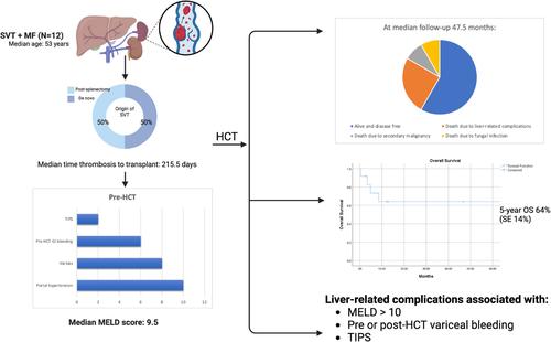
Half of the patients developed SVT following splenectomy, while the other half developed de novo SVT. Three developed SVT within 100 days pre-HCT, all post-splenectomy. The median time from thrombosis to transplant was 215.5 (range: 41–4748) days. The median Yerdel score, describing the extent of thrombosis and risk of complication, was 3 (range: 1–3). Six (50%) patients had chronic SVT and were not anticoagulated pre-HCT. The remaining six (50%) received anticoagulation (DOAC or enoxaparin) for acute thrombosis, with a median duration of 180 days (95–1950). Three (42.8%) continued anticoagulation peri-HCT after platelet recovery. Further information is seen in Table S2.

With regards to liver-related characteristics, the median MELD score was 9.5 (range: 8–17). Most patients had radiologic evidence of PHT (n = 10; 83.3%) or varices (n = 8; 66.6%). Of those with varices, six (75%) had pre-HCT variceal bleeding. Two had pre-emptive banding of varices, and four received beta blocker prophylaxis. Two patients required pre-HCT intervention with transjugular intrahepatic portosystemic shunt (TIPS) and portocaval shunt. No biopsy-proven cirrhosis or iron overload was found pre-HCT, though biopsies were only performed in 4/12 patients. No alternate causes of liver dysfunction were identified. Further information is seen in Table S3.

At Day 100, 11 (92%) patients were alive and disease-free. At a median follow up of 47.5 months, seven (66.6%) were alive and disease-free. Five-year overall survival was 64% (SE 14%). Five patients died: one from secondary malignancy (Day 2315), one from fungal infection related to poor graft function (Day 148), although recurrent gastrointestinal (GI) bleeding and ascites contributed to their decline, and three from direct liver-related complications (veno-occlusive disease [VOD] and intracranial hemorrhage, Day 12; liver failure on Day 103 and liver failure on 260).

For those patients who died from direct liver-related complications: the first patient (MELD 15, pre-HCT variceal bleeding requiring banding) developed severe VOD at Day 4 post-HCT and died of intracranial hemorrhage at Day 12. The second patient (MELD 10, pre-HCT variceal bleeding requiring banding) suffered liver failure following TIPS placement for portal and splenic vein thrombus at Day 62, leading to death on Day 103. The third patient (MELD 13) developed symptomatic ascites post-HCT with imaging revealing acute hepatic vein thrombus (BCS) and cavernous portal vein thrombus requiring TIPS placement on Day 95. She developed progressive liver failure and GI bleeding, dying at Day 260. Overall, liver-related complications were associated with pre-HCT MELD scores > 10, pre- or post-HCT variceal bleeding, TIPS placement, and failure of recanalization on post-HCT imaging.

Non-fatal liver-related complications included liver toxicity and progressive thrombosis. Grade 1 hyperbilirubinemia (n = 3; 25%) and Grade 1 transaminitis (n = 2; 17%) resolved without intervention by Day 100. Five patients (42%) developed long-term sequelae of PHT at a median of 146.5 days (range: 72–3304). These included decompensated ascites (n = 3; 33.3%), variceal bleeding requiring endoscopic management (n = 4; 33.3%), and encephalopathy in the setting of previous TIPS (n = 2; 16%). Median spleen size was 24 cm, and 75% had not undergone splenectomy. Recurrent GI bleeding post-HCT (median 76 days, range: 3–3323) occurred in patients with pre-HCT esophageal varices and no anticoagulation, despite beta blocker prophylaxis. New venous thromboembolism (VTE) was noted in four patients post-HCT, including three (75%) occurring while off anticoagulation. Three (75%) of these events occurred outside the splanchnic system (deep veins of the limbs and pulmonary embolism). Recanalization of SVT occurred in only 16% of patients.

Despite this, some patients had encouraging clinical outcomes at extended follow up. Patient 8 (MELD 8) experienced recurrent variceal bleeding and ascites requiring paracentesis up to 9 years post-HCT, but was alive and clinically stable at 12.5 years post-HCT with long-term hepatology follow-up and diuretic therapy. Patient 6 (MELD 9) developed increasing varices and splenomegaly post-HCT but remained disease-free with no liver toxicity 2577 days post-HCT.

This case series indicates that MF patients with pre-existing SVT can successfully undergo HCT, although they face liver and thrombosis-associated morbidity and mortality. Optimal management of SVT, particularly in the peri-HCT period, is not well studied. However, the rate of recurrent thrombosis in this setting is increased [2]. While anticoagulation in acute thrombosis improves recanalization and reduces PHT, it may increase bleeding, particularly in the setting of varices. Consideration of beta blockade or preemptive banding may mitigate this risk, although this has not been validated in MF. Chronic SVT rarely recanalizes, so life-long anticoagulation is generally recommended to reduce risk of recurrence. In our cohort, long-term anticoagulation, including peri-HCT, was not associated with bleeding, though the decision to continue anticoagulation peri-HCT should be individualized. Results with interventions such as TIPS, thrombolysis, and liver transplantation are variable in non-HCT patients. Portosystemic shunting shows a survival benefit in BCS patients, but its benefits in SVT remain unclear [3].

Management of splenomegaly pre- and peri-HCT is challenging. Splenectomy is often used in an attempt to reverse PHT and improve engraftment, however post-operative SVT rates are as high as 55% with laparoscopic approaches [4]. Morbidity may be significant, including intestinal ischemia and even death. Risk factors for SVT development include splenic weight, co-existent MPN, splenic/portal vein diameter, low white blood counts, and anatomic variation [5]. Half of the patients in our cohort developed SVT following splenectomy, but splenectomy itself did not appear to be associated with long-term morbidity.

MF patients are at risk of post-HCT hepatotoxicity due to iron overload, extramedullary hematopoiesis, and pre-existing PHT. Wong et al. found an increased risk of VOD in patients undergoing HCT for MF, with an association between SVT and hyperbilirubinemia [1]. In contrast, our data showed no persistent liver dysfunction after Day 100, suggesting that with appropriate patient selection, hepatic function is not compromised. One patient died due to complications of VOD, indicating the need for careful surveillance in the post-HCT period.

PHT is a common complication of MF, with potential etiologies including splenomegaly, PVT, extramedullary hematopoiesis and recently, sinusoidal fibrosis [1]. Resultant varices increase the risk of hemorrhage and ascites, leading to long-term morbidity. Thrombosis often contributes, with nearly half of non-HCT patients with JAK2 V617F-associated non-cirrhotic PVT developing PHT during follow-up [6]. In our cohort, sequelae of PHT were evident at Day 100, with persistent morbidity evident even after 12 years in some patients. Liver-related variables such as bilirubin, albumin, and INR, MELD score (> 10) and non-recanalization pre-HCT were key prognostic indicators in this cohort. Other high-risk clinical characteristics, including pre-HCT variceal bleeding, appear to predict similar bleeding post-HCT, regardless of intervention. Pre-HCT optimization, such as variceal banding, may improve outcomes, although this is unclear. All patients undergoing HCT workup should have dedicated imaging for PHT, endoscopic evaluation for variceal treatment and long-term multidisciplinary follow up.

Our study, although limited by its small patient cohort, retrospective design, and the lack of representation of BCS or hepatic vein thrombus, suggests that HCT is feasible and curative for selected patients with SVT with low early and overall mortality. However, the long-term sequelae of PHT remain a concern. Factors such as variceal bleeding and MELD score > 10 appear to increase the risk of future morbidity and mortality related to PHT. Multidisciplinary pre-HCT evaluation including dedicated imaging for PHT, screening and pre-emptive treatment of varices, and long-term follow up contribute to successful outcomes in this patient population. Larger studies are needed to validate these results and refine risk stratification for patients with SVT undergoing HCT.

Abstract Image

求助全文
约1分钟内获得全文 求助全文
来源期刊
CiteScore
15.70
自引率
3.90%
发文量
363
审稿时长
3-6 weeks
期刊介绍: The American Journal of Hematology offers extensive coverage of experimental and clinical aspects of blood diseases in humans and animal models. The journal publishes original contributions in both non-malignant and malignant hematological diseases, encompassing clinical and basic studies in areas such as hemostasis, thrombosis, immunology, blood banking, and stem cell biology. Clinical translational reports highlighting innovative therapeutic approaches for the diagnosis and treatment of hematological diseases are actively encouraged.The American Journal of Hematology features regular original laboratory and clinical research articles, brief research reports, critical reviews, images in hematology, as well as letters and correspondence.
×
引用
GB/T 7714-2015
复制
MLA
复制
APA
复制
导出至
BibTeX EndNote RefMan NoteFirst NoteExpress
×
提示
您的信息不完整,为了账户安全,请先补充。
现在去补充
×
提示
您因"违规操作"
具体请查看互助需知
我知道了
×
提示
确定
请完成安全验证×
copy
已复制链接
快去分享给好友吧!
我知道了
右上角分享
点击右上角分享
0
联系我们:info@booksci.cn Book学术提供免费学术资源搜索服务,方便国内外学者检索中英文文献。致力于提供最便捷和优质的服务体验。 Copyright © 2023 布克学术 All rights reserved.
京ICP备2023020795号-1
ghs 京公网安备 11010802042870号
Book学术文献互助
Book学术文献互助群
群 号:481959085
Book学术官方微信