Multiple loci for foveolar vision in macaque monkey visual cortex
IF 21.2
1区 医学
Q1 NEUROSCIENCES
引用次数: 0
Abstract
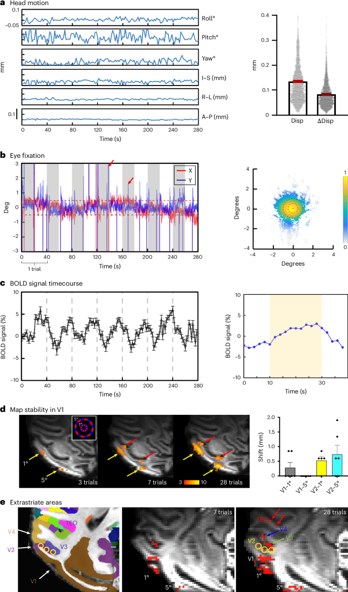
In humans and nonhuman primates, the central 1° of vision is processed by the foveola, a retinal structure that comprises a high density of photoreceptors and is crucial for primate-specific high-acuity vision, color vision and gaze-directed visual attention. Here, we developed high-spatial-resolution ultrahigh-field 7T functional magnetic resonance imaging methods for functional mapping of the foveolar visual cortex in awake monkeys. In the ventral pathway (visual areas V1–V4 and the posterior inferior temporal cortex), viewing of a small foveolar spot elicits a ring of multiple (eight) foveolar representations per hemisphere. This ring surrounds an area called the ‘foveolar core’, which is populated by millimeter-scale functional domains sensitive to fine stimuli and high spatial frequencies, consistent with foveolar visual acuity, color and achromatic information and motion. Thus, this elaborate rerepresentation of central vision coupled with a previously unknown foveolar core area signifies a cortical specialization for primate foveation behaviors. The retinal foveola in the primate eye is critical for seeing fine details, color, text and faces. Using ultrahigh-field functional magnetic resonance imaging, Qian et al discover that there is a highly specialized cortical brain region for processing foveolar information.


猕猴视觉皮层中央窝视觉的多个位点
在人类和非人类灵长类动物中,中央1°的视觉由中央凹处理,中央凹是一种视网膜结构,包含高密度的光感受器,对灵长类动物特有的高敏度视觉、色觉和凝视导向的视觉注意至关重要。本研究利用高空间分辨率的超高场7T功能磁共振成像技术,对清醒猕猴的中央凹视觉皮层进行功能定位。在腹侧通路(V1-V4视觉区和后下颞叶皮层),观察到一个小的中央凹点会在每个半球产生多个(8)个中央凹表征环。这个环围绕着一个被称为“中央焦核”的区域,该区域由毫米级的功能域组成,这些功能域对精细刺激和高空间频率敏感,与中央焦的视觉敏锐度、颜色和消色差信息和运动一致。因此,这种复杂的中央视觉再现加上先前未知的中央窝核心区表明灵长类动物中央窝行为的皮层特化。
本文章由计算机程序翻译,如有差异,请以英文原文为准。
求助全文
约1分钟内获得全文
求助全文
来源期刊

Nature neuroscience
医学-神经科学
CiteScore
38.60
自引率
1.20%
发文量
212
审稿时长
1 months
期刊介绍:
Nature Neuroscience, a multidisciplinary journal, publishes papers of the utmost quality and significance across all realms of neuroscience. The editors welcome contributions spanning molecular, cellular, systems, and cognitive neuroscience, along with psychophysics, computational modeling, and nervous system disorders. While no area is off-limits, studies offering fundamental insights into nervous system function receive priority.
The journal offers high visibility to both readers and authors, fostering interdisciplinary communication and accessibility to a broad audience. It maintains high standards of copy editing and production, rigorous peer review, rapid publication, and operates independently from academic societies and other vested interests.
In addition to primary research, Nature Neuroscience features news and views, reviews, editorials, commentaries, perspectives, book reviews, and correspondence, aiming to serve as the voice of the global neuroscience community.
 求助内容:
求助内容: 应助结果提醒方式:


