Nur Amelia Bachtiar, Bachtiar Murtala, Mirna Muis, Muhammad I Ilyas, Hamzaini Bin Abdul Hamid, Suryani As'ad, Jumraini Tammasse, Audry Devisanty Wuysang, Gita Vita Soraya
{"title":"Non-Contrast MRI Sequences for Ischemic Stroke: A Concise Overview for Clinical Radiologists.","authors":"Nur Amelia Bachtiar, Bachtiar Murtala, Mirna Muis, Muhammad I Ilyas, Hamzaini Bin Abdul Hamid, Suryani As'ad, Jumraini Tammasse, Audry Devisanty Wuysang, Gita Vita Soraya","doi":"10.2147/VHRM.S474143","DOIUrl":null,"url":null,"abstract":"<p><p>Ischemic stroke is the second leading cause of mortality and morbidity worldwide. Due to the urgency of implementing immediate therapy, acute stroke necessitates prompt diagnosis. The current gold standards for vascular imaging in stroke include computed tomography angiography (CTA), digital subtraction angiography (DSA) and magnetic resonance angiography (MRA). However, the contrast agents used in these methods can be costly and pose risks for patients with renal impairment or allergies. The aim of this paper is to provide a comprehensive overview of current MRI techniques and sequences for evaluating ischemic stroke, emphasizing the importance of non-contrast options and their clinical implications for radiologists in the diagnosis and management of ischemic stroke. Standard MRI sequences-such as T1-weighted imaging (T1WI), T2-weighted imaging (T2WI), fluid-attenuated inversion recovery (FLAIR), diffusion-weighted imaging (DWI), DWI-FLAIR mismatch, and apparent diffusion coefficient (ADC)-are essential for determining infarct location, volume, and age. Additionally, incorporating susceptibility-weighted imaging (SWI) sequence aids in identifying signs of hemorrhagic transformation within the infarcted region. Advanced techniques like arterial spin labeling (ASL) can serve as a non-contrast alternative for mapping cerebral blood flow (CBF) and allowing for comparison between infarcted and healthy brain areas. Adding ASL to the routine sequence allows ASL-DWI mismatch analysis that is useful for quantifying salvageable tissue volume and facilitate timely recanalization, while time-of-flight (TOF) MRA and magnetic resonance venography (MRV) help assess venous thrombosis, stenosis, or arterial occlusions. Finally, MR spectroscopy can provide insights into critical brain metabolites, including N-acetylaspartate (NAA), and lactate (Lac) to determine patient prognosis. Current MRI technology provides a myriad of sequence options for the comprehensive evaluation of ischemic stroke without the need for contrast material. A thorough understanding of the advantages and limitations of each sequence is crucial for its optimal implementation in diagnosis and treatment.</p>","PeriodicalId":23597,"journal":{"name":"Vascular Health and Risk Management","volume":"20 ","pages":"521-531"},"PeriodicalIF":2.8000,"publicationDate":"2024-11-26","publicationTypes":"Journal Article","fieldsOfStudy":null,"isOpenAccess":false,"openAccessPdf":"https://www.ncbi.nlm.nih.gov/pmc/articles/PMC11608002/pdf/","citationCount":"0","resultStr":null,"platform":"Semanticscholar","paperid":null,"PeriodicalName":"Vascular Health and Risk Management","FirstCategoryId":"1085","ListUrlMain":"https://doi.org/10.2147/VHRM.S474143","RegionNum":0,"RegionCategory":null,"ArticlePicture":[],"TitleCN":null,"AbstractTextCN":null,"PMCID":null,"EPubDate":"2024/1/1 0:00:00","PubModel":"eCollection","JCR":"Q2","JCRName":"PERIPHERAL VASCULAR DISEASE","Score":null,"Total":0}
引用次数: 0
Abstract
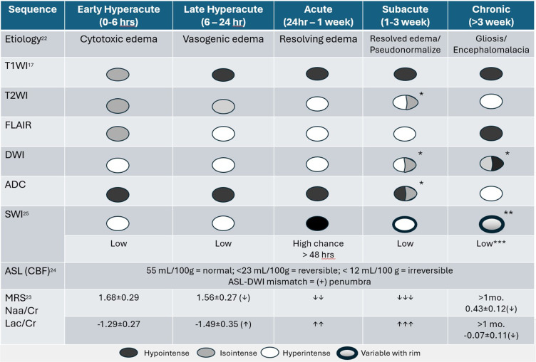
Ischemic stroke is the second leading cause of mortality and morbidity worldwide. Due to the urgency of implementing immediate therapy, acute stroke necessitates prompt diagnosis. The current gold standards for vascular imaging in stroke include computed tomography angiography (CTA), digital subtraction angiography (DSA) and magnetic resonance angiography (MRA). However, the contrast agents used in these methods can be costly and pose risks for patients with renal impairment or allergies. The aim of this paper is to provide a comprehensive overview of current MRI techniques and sequences for evaluating ischemic stroke, emphasizing the importance of non-contrast options and their clinical implications for radiologists in the diagnosis and management of ischemic stroke. Standard MRI sequences-such as T1-weighted imaging (T1WI), T2-weighted imaging (T2WI), fluid-attenuated inversion recovery (FLAIR), diffusion-weighted imaging (DWI), DWI-FLAIR mismatch, and apparent diffusion coefficient (ADC)-are essential for determining infarct location, volume, and age. Additionally, incorporating susceptibility-weighted imaging (SWI) sequence aids in identifying signs of hemorrhagic transformation within the infarcted region. Advanced techniques like arterial spin labeling (ASL) can serve as a non-contrast alternative for mapping cerebral blood flow (CBF) and allowing for comparison between infarcted and healthy brain areas. Adding ASL to the routine sequence allows ASL-DWI mismatch analysis that is useful for quantifying salvageable tissue volume and facilitate timely recanalization, while time-of-flight (TOF) MRA and magnetic resonance venography (MRV) help assess venous thrombosis, stenosis, or arterial occlusions. Finally, MR spectroscopy can provide insights into critical brain metabolites, including N-acetylaspartate (NAA), and lactate (Lac) to determine patient prognosis. Current MRI technology provides a myriad of sequence options for the comprehensive evaluation of ischemic stroke without the need for contrast material. A thorough understanding of the advantages and limitations of each sequence is crucial for its optimal implementation in diagnosis and treatment.
期刊介绍:
An international, peer-reviewed journal of therapeutics and risk management, focusing on concise rapid reporting of clinical studies on the processes involved in the maintenance of vascular health; the monitoring, prevention, and treatment of vascular disease and its sequelae; and the involvement of metabolic disorders, particularly diabetes. In addition, the journal will also seek to define drug usage in terms of ultimate uptake and acceptance by the patient and healthcare professional.

 求助内容:
求助内容: 应助结果提醒方式:


