Zidao Wang, Yuehuan Li, Christian L Andersen, Ahmed E El Zowalaty, Jonathan M Hancock, Taylor E Martin, Elizabeth W Howerth, Suvitha Viswanathan, Haeyeun Byun, Xiaoqin Ye
{"title":"Exogenous estrogen partially rescues progesterone deficiency and autophagosome enlargement in <i>Mcoln1</i> <sup>-/-</sup> mouse model with lysosomal storage disorder.","authors":"Zidao Wang, Yuehuan Li, Christian L Andersen, Ahmed E El Zowalaty, Jonathan M Hancock, Taylor E Martin, Elizabeth W Howerth, Suvitha Viswanathan, Haeyeun Byun, Xiaoqin Ye","doi":"10.1097/RD9.0000000000000109","DOIUrl":null,"url":null,"abstract":"<p><strong>Objective: </strong>Female <i>Mcoln1</i> <sup>-/-</sup> mice exhibit progressive progesterone (P4) deficiency, luteal cell degeneration, and premature embryo implantation failure at 5 months old. We attempted to rescue embryo implantation in non-virgin <i>Mcoln1</i> <sup>-/-</sup> mice (5-6 months old) with exogenous P4 treatment on days 1.5 post-coitum (D1.5), D2.5, and D3.5, and observed partially restored luteal cell morphology on D4.5, but unexpectedly found 17β-estradiol (E<sub>2</sub>) contamination in the P4 working solution. In this study, we aim to investigate exogenous P4 and/or E<sub>2</sub> for the partial recovery of luteal cell morphology in infertile <i>Mcoln1</i> <sup><i>-/-</i></sup> mice.</p><p><strong>Methods: </strong>Control and non-virgin <i>Mcoln1</i> <sup>-/-</sup> mice (5-6 months old) were treated with newly ordered vehicle, P4, E<sub>2</sub>, or P4 + E<sub>2</sub> on D1.5 and D2.5 and dissected on D3.5 for P4 and E<sub>2</sub> measurements, ovary histology, immunofluorescence, lipid droplet staining, and transmission electron microscopy.</p><p><strong>Results: </strong>E<sub>2</sub> treatment significantly increased serum P4 levels in D3.5 <i>Mcoln1</i> <sup>-/-</sup> mice. E<sub>2</sub> and P4 + E<sub>2</sub> treatments, but not P4 treatment alone, largely improved the morphology of D3.5 <i>Mcoln1</i> <sup>-/-</sup> corpora lutea, indicated by a more contiguous web-like collagen IV expression pattern, increased heat shock protein 60 expression, and reduced accumulation of large lipid droplets. Transmission electron microscopy revealed extremely enlarged autophagosomes and lipid droplets, lysosomes with lamellar structures, and mitochondria with reduced cristae in vehicle-treated D3.5 <i>Mcoln1</i> <sup>-/-</sup> luteal cells, while in E<sub>2</sub>-treated D3.5 <i>Mcoln1</i> <sup>-/-</sup> luteal cells, extremely enlarged autophagosomes and lipid droplets were reduced, indicating improved luteal cell ultrastructure.</p><p><strong>Conclusion: </strong>These findings reveal protective effects of high levels of exogenous E<sub>2</sub> on P4 production and lysosomal function in <i>Mcoln1</i> <sup>-/-</sup> luteal cells.</p>","PeriodicalId":20959,"journal":{"name":"Reproductive and Developmental Medicine","volume":"8 4","pages":"197-205"},"PeriodicalIF":0.7000,"publicationDate":"2024-12-01","publicationTypes":"Journal Article","fieldsOfStudy":null,"isOpenAccess":false,"openAccessPdf":"https://www.ncbi.nlm.nih.gov/pmc/articles/PMC11608623/pdf/","citationCount":"0","resultStr":null,"platform":"Semanticscholar","paperid":null,"PeriodicalName":"Reproductive and Developmental Medicine","FirstCategoryId":"3","ListUrlMain":"https://doi.org/10.1097/RD9.0000000000000109","RegionNum":4,"RegionCategory":"医学","ArticlePicture":[],"TitleCN":null,"AbstractTextCN":null,"PMCID":null,"EPubDate":"2024/9/6 0:00:00","PubModel":"Epub","JCR":"Q4","JCRName":"OBSTETRICS & GYNECOLOGY","Score":null,"Total":0}
引用次数: 0
Abstract
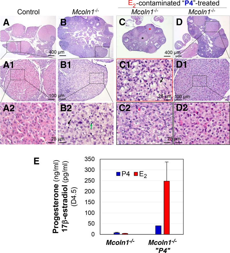
Objective: Female Mcoln1-/- mice exhibit progressive progesterone (P4) deficiency, luteal cell degeneration, and premature embryo implantation failure at 5 months old. We attempted to rescue embryo implantation in non-virgin Mcoln1-/- mice (5-6 months old) with exogenous P4 treatment on days 1.5 post-coitum (D1.5), D2.5, and D3.5, and observed partially restored luteal cell morphology on D4.5, but unexpectedly found 17β-estradiol (E2) contamination in the P4 working solution. In this study, we aim to investigate exogenous P4 and/or E2 for the partial recovery of luteal cell morphology in infertile Mcoln1-/- mice.
Methods: Control and non-virgin Mcoln1-/- mice (5-6 months old) were treated with newly ordered vehicle, P4, E2, or P4 + E2 on D1.5 and D2.5 and dissected on D3.5 for P4 and E2 measurements, ovary histology, immunofluorescence, lipid droplet staining, and transmission electron microscopy.
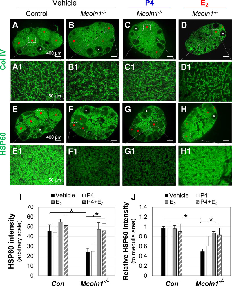
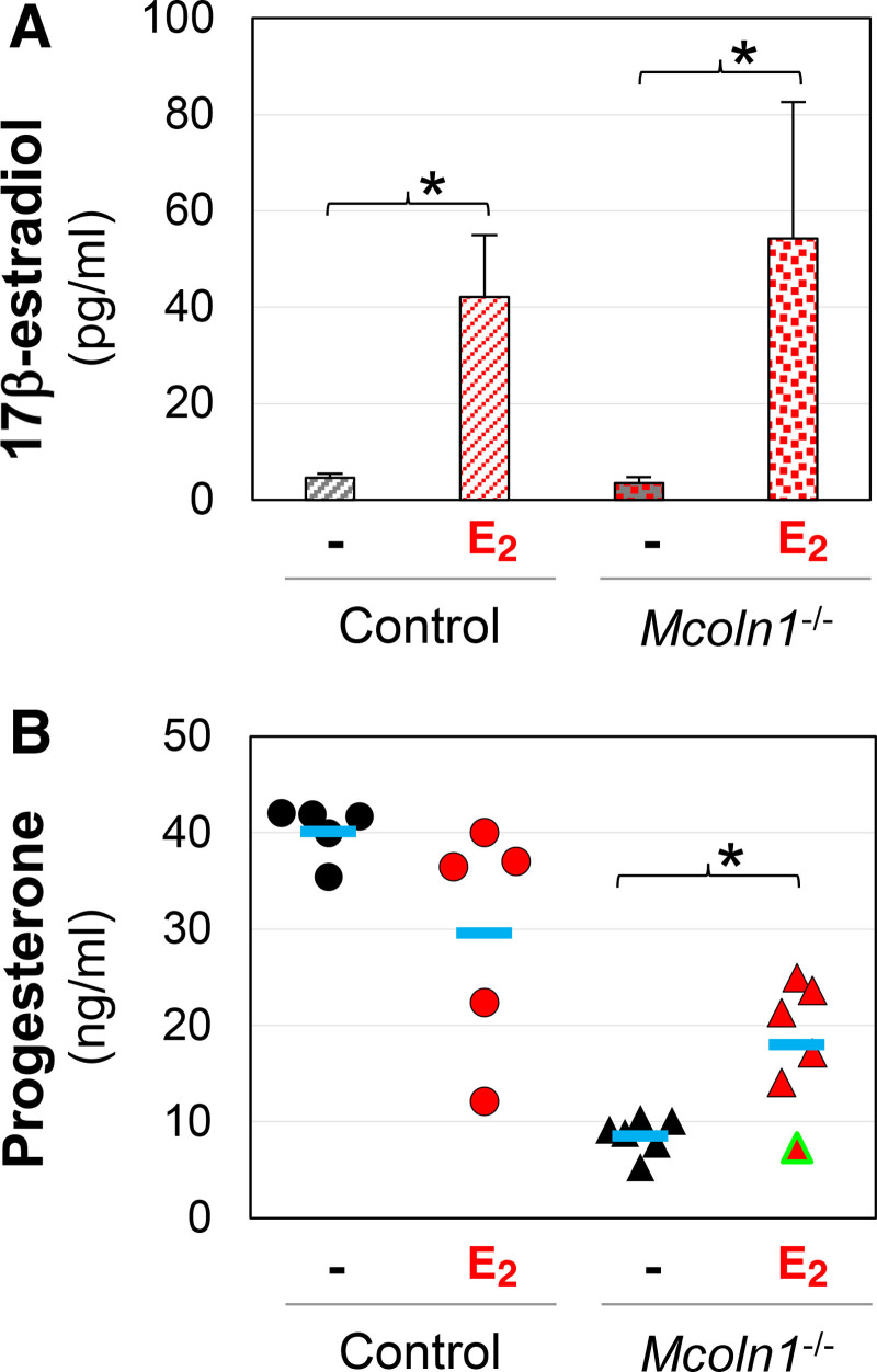
Results: E2 treatment significantly increased serum P4 levels in D3.5 Mcoln1-/- mice. E2 and P4 + E2 treatments, but not P4 treatment alone, largely improved the morphology of D3.5 Mcoln1-/- corpora lutea, indicated by a more contiguous web-like collagen IV expression pattern, increased heat shock protein 60 expression, and reduced accumulation of large lipid droplets. Transmission electron microscopy revealed extremely enlarged autophagosomes and lipid droplets, lysosomes with lamellar structures, and mitochondria with reduced cristae in vehicle-treated D3.5 Mcoln1-/- luteal cells, while in E2-treated D3.5 Mcoln1-/- luteal cells, extremely enlarged autophagosomes and lipid droplets were reduced, indicating improved luteal cell ultrastructure.
Conclusion: These findings reveal protective effects of high levels of exogenous E2 on P4 production and lysosomal function in Mcoln1-/- luteal cells.
 求助内容:
求助内容: 应助结果提醒方式:


