Anatomical Landmarks of the Facial Artery and Vein for Intraoral Anastomosis: A Cadaveric Study

IF 1.5 3区 医学 Q3 SURGERY
Microsurgery Pub Date : 2024-11-29 DOI:10.1002/micr.70004
Kengo Nakatsuka, Tomoyuki Yano, Takuya Omotehara, Shinichi Kawata, Masahiro Itoh
{"title":"Anatomical Landmarks of the Facial Artery and Vein for Intraoral Anastomosis: A Cadaveric Study","authors":"Kengo Nakatsuka,&nbsp;Tomoyuki Yano,&nbsp;Takuya Omotehara,&nbsp;Shinichi Kawata,&nbsp;Masahiro Itoh","doi":"10.1002/micr.70004","DOIUrl":null,"url":null,"abstract":"<div>\n \n \n <section>\n \n <h3> Background</h3>\n \n <p>Intraoral anastomosis is a widely used technique for microvascular alveolar ridge augmentation and midface reconstruction. However, the predictable anatomical positioning of facial structures, such as the vessels, parotid duct, and facial nerve in the buccal region, has remained unclear. Therefore, we aimed to obtain the anatomical characteristics of these locations to establish surgical landmarks for the intraoral anastomosis of facial vessels.</p>\n </section>\n \n <section>\n \n <h3> Methods</h3>\n \n <p>A total of 26 sides from 13 formaldehyde-fixed cadavers approximately a month after fixation with a mean age at death of 86.6 ± 11.2 years (range: 55–104 years) were anatomically examined. Facial vessels, nerves, and the parotid duct were dissected intraorally. From the oral cavity side, the X-axis was defined as the line from the labial commissure to the lowest point of the intertragic notch.</p>\n </section>\n \n <section>\n \n <h3> Results</h3>\n \n <p>From the oral cavity side, all branches of the facial nerve were found under the facial artery and vein. The positioning order along the X-axis was the facial artery, vein, and parotid duct exit. The facial artery was 21.3 ± 2.2 mm and the facial vein was 39.2 ± 2.7 mm from the labial commissure. Ninety-two percent of facial veins were found within 15–20 mm of the facial artery on the X-axis. The parotid duct exit was 46.8 ± 2.0 mm from the labial commissure. In the buccal region, the vessel calibers of the facial artery and vein were 1.8 ± 0.2 and 2.1 ± 0.2 mm, respectively.</p>\n </section>\n \n <section>\n \n <h3> Conclusion</h3>\n \n <p>Knowledge of the anatomical relations among the facial artery, vein, parotid duct, and facial nerve from the oral cavity side can enhance the safety and efficacy of midface reconstruction surgeries involving intraoral anastomosis procedures.</p>\n </section>\n </div>","PeriodicalId":18600,"journal":{"name":"Microsurgery","volume":"45 1","pages":""},"PeriodicalIF":1.5000,"publicationDate":"2024-11-29","publicationTypes":"Journal Article","fieldsOfStudy":null,"isOpenAccess":false,"openAccessPdf":"https://onlinelibrary.wiley.com/doi/epdf/10.1002/micr.70004","citationCount":"0","resultStr":null,"platform":"Semanticscholar","paperid":null,"PeriodicalName":"Microsurgery","FirstCategoryId":"3","ListUrlMain":"https://onlinelibrary.wiley.com/doi/10.1002/micr.70004","RegionNum":3,"RegionCategory":"医学","ArticlePicture":[],"TitleCN":null,"AbstractTextCN":null,"PMCID":null,"EPubDate":"","PubModel":"","JCR":"Q3","JCRName":"SURGERY","Score":null,"Total":0}
引用次数: 0

Abstract

Background

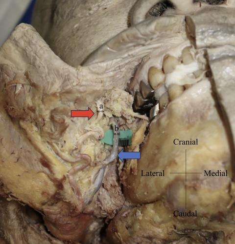
Intraoral anastomosis is a widely used technique for microvascular alveolar ridge augmentation and midface reconstruction. However, the predictable anatomical positioning of facial structures, such as the vessels, parotid duct, and facial nerve in the buccal region, has remained unclear. Therefore, we aimed to obtain the anatomical characteristics of these locations to establish surgical landmarks for the intraoral anastomosis of facial vessels.

Methods

A total of 26 sides from 13 formaldehyde-fixed cadavers approximately a month after fixation with a mean age at death of 86.6 ± 11.2 years (range: 55–104 years) were anatomically examined. Facial vessels, nerves, and the parotid duct were dissected intraorally. From the oral cavity side, the X-axis was defined as the line from the labial commissure to the lowest point of the intertragic notch.

Results

From the oral cavity side, all branches of the facial nerve were found under the facial artery and vein. The positioning order along the X-axis was the facial artery, vein, and parotid duct exit. The facial artery was 21.3 ± 2.2 mm and the facial vein was 39.2 ± 2.7 mm from the labial commissure. Ninety-two percent of facial veins were found within 15–20 mm of the facial artery on the X-axis. The parotid duct exit was 46.8 ± 2.0 mm from the labial commissure. In the buccal region, the vessel calibers of the facial artery and vein were 1.8 ± 0.2 and 2.1 ± 0.2 mm, respectively.

Conclusion

Knowledge of the anatomical relations among the facial artery, vein, parotid duct, and facial nerve from the oral cavity side can enhance the safety and efficacy of midface reconstruction surgeries involving intraoral anastomosis procedures.
Abstract Image

求助全文
约1分钟内获得全文 求助全文
来源期刊
Microsurgery
Microsurgery 医学-外科
CiteScore
3.80
自引率
19.00%
发文量
128
审稿时长
4-8 weeks
期刊介绍: Microsurgery is an international and interdisciplinary publication of original contributions concerning surgery under microscopic magnification. Microsurgery publishes clinical studies, research papers, invited articles, relevant reviews, and other scholarly works from all related fields including orthopaedic surgery, otolaryngology, pediatric surgery, plastic surgery, urology, and vascular surgery.
×
引用
GB/T 7714-2015
复制
MLA
复制
APA
复制
导出至
BibTeX EndNote RefMan NoteFirst NoteExpress
×
提示
您的信息不完整,为了账户安全,请先补充。
现在去补充
×
提示
您因"违规操作"
具体请查看互助需知
我知道了
×
提示
确定
请完成安全验证×
copy
已复制链接
快去分享给好友吧!
我知道了
右上角分享
点击右上角分享
0
联系我们:info@booksci.cn Book学术提供免费学术资源搜索服务,方便国内外学者检索中英文文献。致力于提供最便捷和优质的服务体验。 Copyright © 2023 布克学术 All rights reserved.
京ICP备2023020795号-1
ghs 京公网安备 11010802042870号
Book学术文献互助
Book学术文献互助群
群 号:481959085
Book学术官方微信