{"title":"Hydrogen Protects Mitochondrial Function by Increasing the Expression of PGC-1α and Ameliorating Myocardial Ischaemia–Reperfusion Injury","authors":"Yue Zuo, Jiawei Wang, Zhexuan Gong, Yulong Wang, Qiang Wang, Xueyang Yang, Fulin Liu, Tongtong Liu","doi":"10.1111/jcmm.70236","DOIUrl":null,"url":null,"abstract":"<p>To investigate the application of H<sub>2</sub> to alleviate cardiac ischaemia–reperfusion (I/R) injury in a PGC-1α-dependent manner. A rat in vitro myocardial I/R injury model was used, Western blot was used to detect the expression levels of apoptosis markers (Bax, cleaved caspase-3, Bcl<sub>2</sub>), inflammatory factors (IL-1β, TNF-α), mitochondrial fission (DRP1, MFF) and mitochondrial fusion (MFN1, MFN2, OPA1). HE staining was used to observe the effect of H<sub>2</sub> on the myocardial tissue structure injured by I/R. Transmission electron microscopy (TEM) was used to observe the changes in the mitochondrial structure of myocardial tissue after I/R injury. Real-time quantitative PCR (qPCR) was used to detect the expression of PGC-1α in the myocardial tissue of rats after I/R injury and H<sub>2</sub> treatment. H<sub>2</sub> increases the expression level of PGC-1α, while the deletion of PGC-1α inhibited the therapeutic effect of H<sub>2</sub>. H<sub>2</sub> can improve the changes of the myocardial tissue and mitochondrial structure caused by I/R injury. H<sub>2</sub> treatment effectively inhibited the inflammatory response, and the loss of PGC-1α could inhibit the therapeutic effect of H<sub>2</sub>. The application of H<sub>2</sub> can alleviate myocardial I/R injury, and the loss of PGC-1α weakens the protective effect of H<sub>2</sub> on the I/R heart.</p>","PeriodicalId":101321,"journal":{"name":"JOURNAL OF CELLULAR AND MOLECULAR MEDICINE","volume":"28 22","pages":""},"PeriodicalIF":5.3000,"publicationDate":"2024-11-27","publicationTypes":"Journal Article","fieldsOfStudy":null,"isOpenAccess":false,"openAccessPdf":"https://onlinelibrary.wiley.com/doi/epdf/10.1111/jcmm.70236","citationCount":"0","resultStr":null,"platform":"Semanticscholar","paperid":null,"PeriodicalName":"JOURNAL OF CELLULAR AND MOLECULAR MEDICINE","FirstCategoryId":"1085","ListUrlMain":"https://onlinelibrary.wiley.com/doi/10.1111/jcmm.70236","RegionNum":0,"RegionCategory":null,"ArticlePicture":[],"TitleCN":null,"AbstractTextCN":null,"PMCID":null,"EPubDate":"","PubModel":"","JCR":"","JCRName":"","Score":null,"Total":0}
引用次数: 0
Abstract
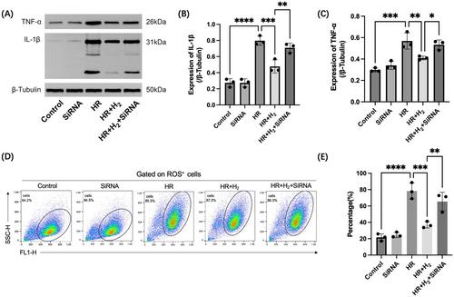
To investigate the application of H2 to alleviate cardiac ischaemia–reperfusion (I/R) injury in a PGC-1α-dependent manner. A rat in vitro myocardial I/R injury model was used, Western blot was used to detect the expression levels of apoptosis markers (Bax, cleaved caspase-3, Bcl2), inflammatory factors (IL-1β, TNF-α), mitochondrial fission (DRP1, MFF) and mitochondrial fusion (MFN1, MFN2, OPA1). HE staining was used to observe the effect of H2 on the myocardial tissue structure injured by I/R. Transmission electron microscopy (TEM) was used to observe the changes in the mitochondrial structure of myocardial tissue after I/R injury. Real-time quantitative PCR (qPCR) was used to detect the expression of PGC-1α in the myocardial tissue of rats after I/R injury and H2 treatment. H2 increases the expression level of PGC-1α, while the deletion of PGC-1α inhibited the therapeutic effect of H2. H2 can improve the changes of the myocardial tissue and mitochondrial structure caused by I/R injury. H2 treatment effectively inhibited the inflammatory response, and the loss of PGC-1α could inhibit the therapeutic effect of H2. The application of H2 can alleviate myocardial I/R injury, and the loss of PGC-1α weakens the protective effect of H2 on the I/R heart.
期刊介绍:
The Journal of Cellular and Molecular Medicine serves as a bridge between physiology and cellular medicine, as well as molecular biology and molecular therapeutics. With a 20-year history, the journal adopts an interdisciplinary approach to showcase innovative discoveries.
It publishes research aimed at advancing the collective understanding of the cellular and molecular mechanisms underlying diseases. The journal emphasizes translational studies that translate this knowledge into therapeutic strategies. Being fully open access, the journal is accessible to all readers.
 求助内容:
求助内容: 应助结果提醒方式:


