Guanrong Ma, Zhiqi Chen, Lanxi Wang, Xiulin Chang, Liaoqiong Fang, Jin Bai
{"title":"Study on the biosafety and targeting efficiency of Escherichia coli outer membrane vesicles in breast tumor","authors":"Guanrong Ma, Zhiqi Chen, Lanxi Wang, Xiulin Chang, Liaoqiong Fang, Jin Bai","doi":"10.1007/s10735-024-10296-0","DOIUrl":null,"url":null,"abstract":"<div><p>To seek out the targeting of <i>Escherichia coli</i> outer membrane vesicles (<i>E. coli</i> OMVs) in breast tumor-bearing mice and their biosafety in healthy mice. Ultrafiltration in conjunction with ultracentrifugation was utilized to concentrate <i>E. coli</i> OMVs, and characterize them. Subcutaneous breast tumors were induced in BALB/c mice to serve as an experimental model, and the biodistribution of <i>E. coli</i> OMVs in both tumor-bearing and healthy mice was monitored using an in vivo fluorescence imaging system. Utilizing frozen sections, the infiltration of <i>E. coli</i> OMVs in tumor tissues was appraised at the 24-hour post-injection. Healthy BALB/c mice were randomly divided into control group and vesicles group. Following the intravenous injection of <i>E. coli</i> OMVs, monitoring encompassed variations in body weight, blood routine indices, serum levels of AST, ALT, and BUN, organ indices (heart, liver, spleen, lung, and kidney), along with tissue histopathology over a 14-day period. The spherical <i>E. coli</i> OMVs had a diameter of (155.8 ± 3.1) nm and exhibited the expression of outer membrane proteins OmpA and OmpC. Upon assessment, it was evident that the <i>E. coli</i> OMVs persisted in the tumor tissues even 24 h post-injection. An evident decrease in the body weight of the vesicles group, distinct from the control group, was observed on the second day after injection (<i>P</i> < 0.001); in contrast, no considerable differences were noted at subsequent time points (<i>P</i> > 0.05). Following the injection, the vesicles group displayed notable reductions in WBC and PLT as relative to the control group (<i>P</i> < 0.0001) on the initial day, however, there were no noteworthy distinctions as opposed to the control group for other hematological indices; No notable variances in hematological indices between the two groups were observed on the seventh and fourteenth day (<i>P</i> > 0.05). Over the 14 days, no substantial differences were observed in the serum levels of BUN, AST, ALT, and organ indices within the vesicles group as opposed to the control group (<i>P</i> > 0.05). Furthermore, there were no obvious abnormal changes in tissue morphology. 0.5 mg/kg of <i>E. coli</i> OMVs can safely and effectively target 4T1 breast tumor in mice.</p></div>","PeriodicalId":650,"journal":{"name":"Journal of Molecular Histology","volume":"56 1","pages":""},"PeriodicalIF":2.9000,"publicationDate":"2024-11-27","publicationTypes":"Journal Article","fieldsOfStudy":null,"isOpenAccess":false,"openAccessPdf":"","citationCount":"0","resultStr":null,"platform":"Semanticscholar","paperid":null,"PeriodicalName":"Journal of Molecular Histology","FirstCategoryId":"99","ListUrlMain":"https://link.springer.com/article/10.1007/s10735-024-10296-0","RegionNum":4,"RegionCategory":"生物学","ArticlePicture":[],"TitleCN":null,"AbstractTextCN":null,"PMCID":null,"EPubDate":"","PubModel":"","JCR":"Q3","JCRName":"CELL BIOLOGY","Score":null,"Total":0}
引用次数: 0
Abstract
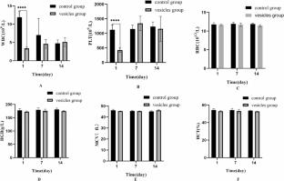
To seek out the targeting of Escherichia coli outer membrane vesicles (E. coli OMVs) in breast tumor-bearing mice and their biosafety in healthy mice. Ultrafiltration in conjunction with ultracentrifugation was utilized to concentrate E. coli OMVs, and characterize them. Subcutaneous breast tumors were induced in BALB/c mice to serve as an experimental model, and the biodistribution of E. coli OMVs in both tumor-bearing and healthy mice was monitored using an in vivo fluorescence imaging system. Utilizing frozen sections, the infiltration of E. coli OMVs in tumor tissues was appraised at the 24-hour post-injection. Healthy BALB/c mice were randomly divided into control group and vesicles group. Following the intravenous injection of E. coli OMVs, monitoring encompassed variations in body weight, blood routine indices, serum levels of AST, ALT, and BUN, organ indices (heart, liver, spleen, lung, and kidney), along with tissue histopathology over a 14-day period. The spherical E. coli OMVs had a diameter of (155.8 ± 3.1) nm and exhibited the expression of outer membrane proteins OmpA and OmpC. Upon assessment, it was evident that the E. coli OMVs persisted in the tumor tissues even 24 h post-injection. An evident decrease in the body weight of the vesicles group, distinct from the control group, was observed on the second day after injection (P < 0.001); in contrast, no considerable differences were noted at subsequent time points (P > 0.05). Following the injection, the vesicles group displayed notable reductions in WBC and PLT as relative to the control group (P < 0.0001) on the initial day, however, there were no noteworthy distinctions as opposed to the control group for other hematological indices; No notable variances in hematological indices between the two groups were observed on the seventh and fourteenth day (P > 0.05). Over the 14 days, no substantial differences were observed in the serum levels of BUN, AST, ALT, and organ indices within the vesicles group as opposed to the control group (P > 0.05). Furthermore, there were no obvious abnormal changes in tissue morphology. 0.5 mg/kg of E. coli OMVs can safely and effectively target 4T1 breast tumor in mice.
期刊介绍:
The Journal of Molecular Histology publishes results of original research on the localization and expression of molecules in animal cells, tissues and organs. Coverage includes studies describing novel cellular or ultrastructural distributions of molecules which provide insight into biochemical or physiological function, development, histologic structure and disease processes.
Major research themes of particular interest include:
- Cell-Cell and Cell-Matrix Interactions;
- Connective Tissues;
- Development and Disease;
- Neuroscience.
Please note that the Journal of Molecular Histology does not consider manuscripts dealing with the application of immunological or other probes on non-standard laboratory animal models unless the results are clearly of significant and general biological importance.
The Journal of Molecular Histology publishes full-length original research papers, review articles, short communications and letters to the editors. All manuscripts are typically reviewed by two independent referees. The Journal of Molecular Histology is a continuation of The Histochemical Journal.

 求助内容:
求助内容: 应助结果提醒方式:


