Wasim Akram, Abul Kalam Najmi, Syed Ehtaishamul Haque
{"title":"Levocabastine ameliorates cyclophosphamide-induced hepatotoxicity in Swiss albino mice: modulation of Nrf2, NF-κB p65, cleaved caspase-3 and TGF-β signaling molecules","authors":"Wasim Akram, Abul Kalam Najmi, Syed Ehtaishamul Haque","doi":"10.1007/s10735-024-10286-2","DOIUrl":null,"url":null,"abstract":"<div><h3>Background</h3><p>Cyclophosphamide (CP)-induced hepatotoxicity is a significant problem in clinical settings. This study aimed to evaluate the protective effect of levocabastine (LEV) on CP-induced hepatotoxicity in Swiss albino mice.</p><h3>Methods and results</h3><p>Mice were given CP (toxic drug) 200 mg/kg, i.p., once on the 7th day, and LEV 50 and 100 µg/kg, i.p., and fenofibrate (FF) 80 mg/kg, p.o., daily for 14 days. On the 15th day, blood and liver samples were collected to assess biological parameters. CP 200 mg/kg caused hepatotoxicity due to oxidative stress, inflammation, apoptosis, and fibrosis as manifested by a reduction in catalase, reduced glutathione (GSH), superoxide dismutase (SOD), and an increase in thiobarbituric acid reactive substance (TBARS), nitrite, tumor necrosis factor-alpha (TNF-α), interleukin-6 (IL-6), transforming growth factor-beta 1 (TGF-β1), interleukin-1β (IL-1β), alkaline phosphatase (ALP), alanine aminotransferase (ALT), aspartate aminotransferase (AST), and gamma-glutamyl transferase (GGT) levels. Cleaved caspase-3 and nuclear factor kappa-B (NF-κB) expression was also increased and nuclear factor erythroid 2-related factor (Nrf2) expression was decreased as confirmed by Immunohistochemical analysis. It also caused histopathological abnormalities and fibrosis as manifested by Hematoxylin-Eosin (H&E) and Masson’s trichrome (MT) staining. These alterations were returned to almost normal when treated with LEV 100 µg/kg and FF 80 mg/kg. Thus, LEV protected CP-induced hepatotoxicity by reversing inflammation, apoptosis, fibrosis, oxidative stress, hepatic injury, and histopathological damages.</p><h3>Conclusion</h3><p>LEV can be helpful as an adjuvant in cancer patients who are on CP treatment, to minimize toxicity. However, its role in in-vivo cancer model is further needed to be confirmed.</p></div>","PeriodicalId":650,"journal":{"name":"Journal of Molecular Histology","volume":"56 1","pages":""},"PeriodicalIF":2.9000,"publicationDate":"2024-11-27","publicationTypes":"Journal Article","fieldsOfStudy":null,"isOpenAccess":false,"openAccessPdf":"","citationCount":"0","resultStr":null,"platform":"Semanticscholar","paperid":null,"PeriodicalName":"Journal of Molecular Histology","FirstCategoryId":"99","ListUrlMain":"https://link.springer.com/article/10.1007/s10735-024-10286-2","RegionNum":4,"RegionCategory":"生物学","ArticlePicture":[],"TitleCN":null,"AbstractTextCN":null,"PMCID":null,"EPubDate":"","PubModel":"","JCR":"Q3","JCRName":"CELL BIOLOGY","Score":null,"Total":0}
引用次数: 0
Abstract
Background
Cyclophosphamide (CP)-induced hepatotoxicity is a significant problem in clinical settings. This study aimed to evaluate the protective effect of levocabastine (LEV) on CP-induced hepatotoxicity in Swiss albino mice.
Methods and results
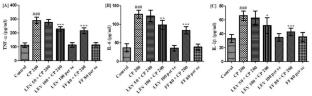
Mice were given CP (toxic drug) 200 mg/kg, i.p., once on the 7th day, and LEV 50 and 100 µg/kg, i.p., and fenofibrate (FF) 80 mg/kg, p.o., daily for 14 days. On the 15th day, blood and liver samples were collected to assess biological parameters. CP 200 mg/kg caused hepatotoxicity due to oxidative stress, inflammation, apoptosis, and fibrosis as manifested by a reduction in catalase, reduced glutathione (GSH), superoxide dismutase (SOD), and an increase in thiobarbituric acid reactive substance (TBARS), nitrite, tumor necrosis factor-alpha (TNF-α), interleukin-6 (IL-6), transforming growth factor-beta 1 (TGF-β1), interleukin-1β (IL-1β), alkaline phosphatase (ALP), alanine aminotransferase (ALT), aspartate aminotransferase (AST), and gamma-glutamyl transferase (GGT) levels. Cleaved caspase-3 and nuclear factor kappa-B (NF-κB) expression was also increased and nuclear factor erythroid 2-related factor (Nrf2) expression was decreased as confirmed by Immunohistochemical analysis. It also caused histopathological abnormalities and fibrosis as manifested by Hematoxylin-Eosin (H&E) and Masson’s trichrome (MT) staining. These alterations were returned to almost normal when treated with LEV 100 µg/kg and FF 80 mg/kg. Thus, LEV protected CP-induced hepatotoxicity by reversing inflammation, apoptosis, fibrosis, oxidative stress, hepatic injury, and histopathological damages.
Conclusion
LEV can be helpful as an adjuvant in cancer patients who are on CP treatment, to minimize toxicity. However, its role in in-vivo cancer model is further needed to be confirmed.
期刊介绍:
The Journal of Molecular Histology publishes results of original research on the localization and expression of molecules in animal cells, tissues and organs. Coverage includes studies describing novel cellular or ultrastructural distributions of molecules which provide insight into biochemical or physiological function, development, histologic structure and disease processes.
Major research themes of particular interest include:
- Cell-Cell and Cell-Matrix Interactions;
- Connective Tissues;
- Development and Disease;
- Neuroscience.
Please note that the Journal of Molecular Histology does not consider manuscripts dealing with the application of immunological or other probes on non-standard laboratory animal models unless the results are clearly of significant and general biological importance.
The Journal of Molecular Histology publishes full-length original research papers, review articles, short communications and letters to the editors. All manuscripts are typically reviewed by two independent referees. The Journal of Molecular Histology is a continuation of The Histochemical Journal.

 求助内容:
求助内容: 应助结果提醒方式:


