A novel three-dimensional method for detailed analysis of RGC central projections under acute ocular hypertension

IF 3 2区 医学 Q1 OPHTHALMOLOGY
Wenhan Lu , Yu Wang , Wei Hu , Xinyi Lin , Xiaoyu Tong , Yi Tian , Yuning Chen , Yicong Wang , Yan Xiao , Hongfang Yang , Yi Feng , Xinghuai Sun
{"title":"A novel three-dimensional method for detailed analysis of RGC central projections under acute ocular hypertension","authors":"Wenhan Lu ,&nbsp;Yu Wang ,&nbsp;Wei Hu ,&nbsp;Xinyi Lin ,&nbsp;Xiaoyu Tong ,&nbsp;Yi Tian ,&nbsp;Yuning Chen ,&nbsp;Yicong Wang ,&nbsp;Yan Xiao ,&nbsp;Hongfang Yang ,&nbsp;Yi Feng ,&nbsp;Xinghuai Sun","doi":"10.1016/j.exer.2024.110157","DOIUrl":null,"url":null,"abstract":"<div><div>Normal perception of visual information relies not only on the quantity and quality of retinal ganglion cells (RGCs), but also on the integrity of the visual pathway, within which RGC central projection predominates. However, the exact changes of RGC central projection under particular pathological conditions remain to be elucidated. Here, we report a whole-brain clearing method modified from iDISCO for 3D visualization of RGC central projection. The CTB-labeled RGC central projection was visualized three-dimensionally with minimized both fluorescence quenching and the time taken. For observation of RGC axonal degeneration pattern under pathological conditions, we took acute ocular hypertension (AOH) as an example. Mice were intracamerally irrigated, and fluorescent signal in brain subregions where RGC axons projected to were quantified. The novel methodology is well-applied for rapid clearing and observation of RGC central projection in C57BL/6J, showing damaged RGC central projection on the AOH side and the most statistically significant degeneration in the superior colliculi (SC). Detailed analysis also revealed a distinct injury pattern among lateral geniculate nuclei (LGN) subregions, with the parvocellular part of the pregeniculate nuclei (PrGPC) being more vulnerable compared with the magnocellular part (PrGMC). The intracranial retrograde labeling of RGC subgroups based on brain damage variation showed PrGPC-projecting RGCs (<em>Plgn</em> RGC) being smaller than PrGMC-projecting RGCs (<em>Mlgn</em> RGC) in size and less in number, yet more vulnerable in terms of degeneration under AOH. Our data revealed the methodology for visualizing selective neuronal vulnerability under AOH, and in the meantime provided novel approach for future mechanisms exploration regarding RGC degeneration.</div></div>","PeriodicalId":12177,"journal":{"name":"Experimental eye research","volume":"250 ","pages":"Article 110157"},"PeriodicalIF":3.0000,"publicationDate":"2024-11-20","publicationTypes":"Journal Article","fieldsOfStudy":null,"isOpenAccess":false,"openAccessPdf":"","citationCount":"0","resultStr":null,"platform":"Semanticscholar","paperid":null,"PeriodicalName":"Experimental eye research","FirstCategoryId":"3","ListUrlMain":"https://www.sciencedirect.com/science/article/pii/S0014483524003798","RegionNum":2,"RegionCategory":"医学","ArticlePicture":[],"TitleCN":null,"AbstractTextCN":null,"PMCID":null,"EPubDate":"","PubModel":"","JCR":"Q1","JCRName":"OPHTHALMOLOGY","Score":null,"Total":0}
引用次数: 0

Abstract

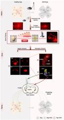
Normal perception of visual information relies not only on the quantity and quality of retinal ganglion cells (RGCs), but also on the integrity of the visual pathway, within which RGC central projection predominates. However, the exact changes of RGC central projection under particular pathological conditions remain to be elucidated. Here, we report a whole-brain clearing method modified from iDISCO for 3D visualization of RGC central projection. The CTB-labeled RGC central projection was visualized three-dimensionally with minimized both fluorescence quenching and the time taken. For observation of RGC axonal degeneration pattern under pathological conditions, we took acute ocular hypertension (AOH) as an example. Mice were intracamerally irrigated, and fluorescent signal in brain subregions where RGC axons projected to were quantified. The novel methodology is well-applied for rapid clearing and observation of RGC central projection in C57BL/6J, showing damaged RGC central projection on the AOH side and the most statistically significant degeneration in the superior colliculi (SC). Detailed analysis also revealed a distinct injury pattern among lateral geniculate nuclei (LGN) subregions, with the parvocellular part of the pregeniculate nuclei (PrGPC) being more vulnerable compared with the magnocellular part (PrGMC). The intracranial retrograde labeling of RGC subgroups based on brain damage variation showed PrGPC-projecting RGCs (Plgn RGC) being smaller than PrGMC-projecting RGCs (Mlgn RGC) in size and less in number, yet more vulnerable in terms of degeneration under AOH. Our data revealed the methodology for visualizing selective neuronal vulnerability under AOH, and in the meantime provided novel approach for future mechanisms exploration regarding RGC degeneration.
Abstract Image

详细分析急性眼压过高症下 RGC 中央投影的新型三维方法
正常的视觉信息感知不仅依赖于视网膜神经节细胞(RGC)的数量和质量,还依赖于视觉通路的完整性,而在视觉通路中,RGC的中心投射占主导地位。然而,RGC中心投射在特定病理条件下的确切变化仍有待阐明。在此,我们报告了一种改良自 iDISCO 的全脑清除方法,用于 RGC 中央投射的三维可视化。在三维观察CTB标记的RGC中心投影时,荧光淬灭和所需时间都降到了最低。为了观察病理条件下RGC轴突变性的模式,我们以急性眼压升高(AOH)为例。对小鼠进行组内灌洗,并对RGC轴突投射到的脑亚区域的荧光信号进行量化。这种新方法适用于快速清除和观察C57BL/6J的RGC中心投射,结果显示AOH一侧的RGC中心投射受损,而上丘脑(SC)的退化在统计学上最为显著。详细分析还显示,外侧膝状核(LGN)亚区域之间存在不同的损伤模式,与大细胞部分(PrGMC)相比,前膝状核的旁细胞部分(PrGPC)更容易受到损伤。根据脑损伤变化对RGC亚群进行的颅内逆行标记显示,PrGPC投射的RGC(Plgn RGC)比PrGMC投射的RGC(Mlgn RGC)体积更小,数量更少,但在AOH作用下更容易发生变性。我们的数据揭示了AOH条件下神经元选择性脆弱性的可视化方法,同时也为未来探索RGC变性的机制提供了新方法。
本文章由计算机程序翻译,如有差异,请以英文原文为准。
求助全文
约1分钟内获得全文 求助全文
来源期刊
Experimental eye research
Experimental eye research 医学-眼科学
CiteScore
6.80
自引率
5.90%
发文量
323
审稿时长
66 days
期刊介绍: The primary goal of Experimental Eye Research is to publish original research papers on all aspects of experimental biology of the eye and ocular tissues that seek to define the mechanisms of normal function and/or disease. Studies of ocular tissues that encompass the disciplines of cell biology, developmental biology, genetics, molecular biology, physiology, biochemistry, biophysics, immunology or microbiology are most welcomed. Manuscripts that are purely clinical or in a surgical area of ophthalmology are not appropriate for submission to Experimental Eye Research and if received will be returned without review.
×
引用
GB/T 7714-2015
复制
MLA
复制
APA
复制
导出至
BibTeX EndNote RefMan NoteFirst NoteExpress
×
提示
您的信息不完整,为了账户安全,请先补充。
现在去补充
×
提示
您因"违规操作"
具体请查看互助需知
我知道了
×
提示
确定
请完成安全验证×
copy
已复制链接
快去分享给好友吧!
我知道了
右上角分享
点击右上角分享
0
联系我们:info@booksci.cn Book学术提供免费学术资源搜索服务,方便国内外学者检索中英文文献。致力于提供最便捷和优质的服务体验。 Copyright © 2023 布克学术 All rights reserved.
京ICP备2023020795号-1
ghs 京公网安备 11010802042870号
Book学术文献互助
Book学术文献互助群
群 号:481959085
Book学术官方微信