Quantification of hepatic fat: evaluation of different magnetic resonance imaging measurement strategies in cases of homogeneous and heterogeneous distribution.
Eloa de Castro Noguerol, Luis Ronan Marquez Ferreira de Souza, Valdair Francisco Muglia, Jorge Elias
{"title":"Quantification of hepatic fat: evaluation of different magnetic resonance imaging measurement strategies in cases of homogeneous and heterogeneous distribution.","authors":"Eloa de Castro Noguerol, Luis Ronan Marquez Ferreira de Souza, Valdair Francisco Muglia, Jorge Elias","doi":"10.1590/0100-3984.2024.0009-en","DOIUrl":null,"url":null,"abstract":"<p><strong>Objective: </strong>To evaluate three different measurements strategies to quantify hepatic steatosis and to investigate the differences between homogeneous and heterogeneous forms of hepatic steatosis.</p><p><strong>Materials and methods: </strong>Retrospective study conducted by magnetic resonance imaging review. We evaluated three different strategies measures for quantification of hepatic steatosis in two matched groups: homogeneous and heterogeneous steatosis. We considered <i>p</i> < 0.05 significance level in all made tests.</p><p><strong>Results: </strong>In heterogeneous steatosis group, the strategy with a region of interest (ROI) of 1 cm<sup>2</sup> to measure the signal intensity in the most altered area showed significant variations in the quantification, while the average of four ROIs of 1 cm<sup>2</sup> or representative target area in axial section did not vary significant. In diffuse hepatic steatosis, any strategy used showed no significant difference. The intraclass correlation coefficient ranged between 0.96 and 0.99, with 95% confidence interval of 0.93-0.99.</p><p><strong>Conclusion: </strong>The quantification of fat liver by magnetic resonance imaging using only one ROI is less representative, especially in heterogeneous steatosis. There was no significant difference between the average of four ROIs strategy and the strategy of representative segmentation area of parenchyma.</p>","PeriodicalId":20842,"journal":{"name":"Radiologia Brasileira","volume":"57 ","pages":"e20240009en"},"PeriodicalIF":0.0000,"publicationDate":"2024-11-18","publicationTypes":"Journal Article","fieldsOfStudy":null,"isOpenAccess":false,"openAccessPdf":"https://www.ncbi.nlm.nih.gov/pmc/articles/PMC11575847/pdf/","citationCount":"0","resultStr":null,"platform":"Semanticscholar","paperid":null,"PeriodicalName":"Radiologia Brasileira","FirstCategoryId":"1085","ListUrlMain":"https://doi.org/10.1590/0100-3984.2024.0009-en","RegionNum":0,"RegionCategory":null,"ArticlePicture":[],"TitleCN":null,"AbstractTextCN":null,"PMCID":null,"EPubDate":"2024/1/1 0:00:00","PubModel":"eCollection","JCR":"Q3","JCRName":"Medicine","Score":null,"Total":0}
引用次数: 0
Abstract
Objective: To evaluate three different measurements strategies to quantify hepatic steatosis and to investigate the differences between homogeneous and heterogeneous forms of hepatic steatosis.
Materials and methods: Retrospective study conducted by magnetic resonance imaging review. We evaluated three different strategies measures for quantification of hepatic steatosis in two matched groups: homogeneous and heterogeneous steatosis. We considered p < 0.05 significance level in all made tests.
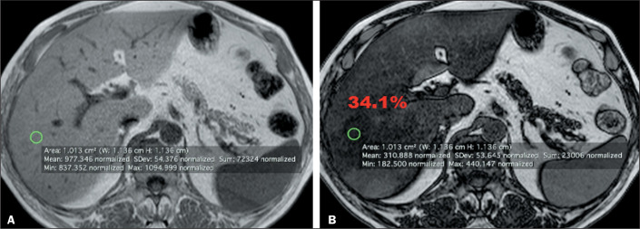
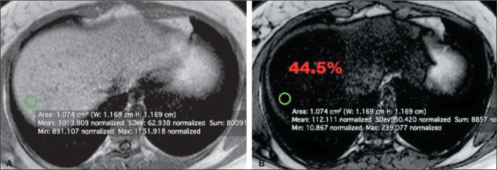
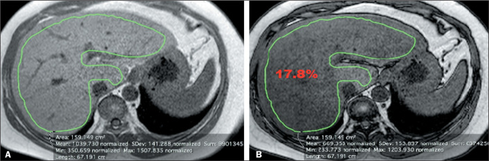
Results: In heterogeneous steatosis group, the strategy with a region of interest (ROI) of 1 cm2 to measure the signal intensity in the most altered area showed significant variations in the quantification, while the average of four ROIs of 1 cm2 or representative target area in axial section did not vary significant. In diffuse hepatic steatosis, any strategy used showed no significant difference. The intraclass correlation coefficient ranged between 0.96 and 0.99, with 95% confidence interval of 0.93-0.99.
Conclusion: The quantification of fat liver by magnetic resonance imaging using only one ROI is less representative, especially in heterogeneous steatosis. There was no significant difference between the average of four ROIs strategy and the strategy of representative segmentation area of parenchyma.

 求助内容:
求助内容: 应助结果提醒方式:


