Stereological Analysis of the Rhesus Monkey Perirhinal and Parahippocampal Cortices

IF 2.3 4区 医学 Q3 NEUROSCIENCES
Justine Villard, Loïc J. Chareyron, Pamela Banta Lavenex, David G. Amaral, Pierre Lavenex
{"title":"Stereological Analysis of the Rhesus Monkey Perirhinal and Parahippocampal Cortices","authors":"Justine Villard,&nbsp;Loïc J. Chareyron,&nbsp;Pamela Banta Lavenex,&nbsp;David G. Amaral,&nbsp;Pierre Lavenex","doi":"10.1002/cne.25684","DOIUrl":null,"url":null,"abstract":"<div>\n \n \n <section>\n \n <p>The perirhinal and parahippocampal cortices are key components of the medial temporal lobe memory system. Despite their essential roles in mnemonic and perceptual functions, there is limited quantitative information regarding their structural characteristics. Here, we implemented design-based stereological techniques to provide estimates of neuron number, neuronal soma size, and volume of the different layers and subdivisions of the perirhinal and parahippocampal cortices in adult macaque monkeys (<i>Macaca mulatta</i>, 5–9 years of age). We found that areas 36r and 36c of the perirhinal cortex and areas TF and TH of the parahippocampal cortex exhibit relatively large superficial layers, which are characteristic of the laminar organization of higher order associational cortices. In contrast, area 35 of the perirhinal cortex exhibits relatively large deep layers. Although neuronal soma size varies between subdivisions and layers, neurons are generally larger in the perirhinal cortex than in the parahippocampal cortex and even larger in the entorhinal cortex. These morphological characteristics are consistent with the hierarchical organization of these cortices within the medial temporal lobe. Comparing data in rats, monkeys, and humans, we found species differences in the relative size of these structures, showing that the perirhinal and parahippocampal cortices have expanded in parallel to the cerebral cortex and may play a greater role in the integration of information in the neocortical–hippocampal loop in primates. Altogether, these normative data provide an essential reference to extrapolate findings from experimental studies in animals and create realistic models of the medial temporal lobe memory system.</p>\n </section>\n </div>","PeriodicalId":15552,"journal":{"name":"Journal of Comparative Neurology","volume":"532 11","pages":""},"PeriodicalIF":2.3000,"publicationDate":"2024-11-17","publicationTypes":"Journal Article","fieldsOfStudy":null,"isOpenAccess":false,"openAccessPdf":"https://onlinelibrary.wiley.com/doi/epdf/10.1002/cne.25684","citationCount":"0","resultStr":null,"platform":"Semanticscholar","paperid":null,"PeriodicalName":"Journal of Comparative Neurology","FirstCategoryId":"3","ListUrlMain":"https://onlinelibrary.wiley.com/doi/10.1002/cne.25684","RegionNum":4,"RegionCategory":"医学","ArticlePicture":[],"TitleCN":null,"AbstractTextCN":null,"PMCID":null,"EPubDate":"","PubModel":"","JCR":"Q3","JCRName":"NEUROSCIENCES","Score":null,"Total":0}
引用次数: 0

Abstract

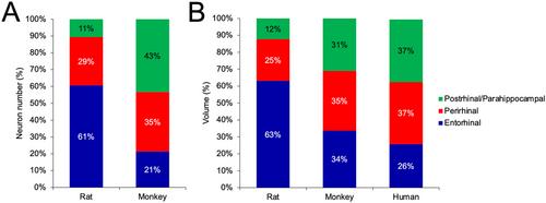
The perirhinal and parahippocampal cortices are key components of the medial temporal lobe memory system. Despite their essential roles in mnemonic and perceptual functions, there is limited quantitative information regarding their structural characteristics. Here, we implemented design-based stereological techniques to provide estimates of neuron number, neuronal soma size, and volume of the different layers and subdivisions of the perirhinal and parahippocampal cortices in adult macaque monkeys (Macaca mulatta, 5–9 years of age). We found that areas 36r and 36c of the perirhinal cortex and areas TF and TH of the parahippocampal cortex exhibit relatively large superficial layers, which are characteristic of the laminar organization of higher order associational cortices. In contrast, area 35 of the perirhinal cortex exhibits relatively large deep layers. Although neuronal soma size varies between subdivisions and layers, neurons are generally larger in the perirhinal cortex than in the parahippocampal cortex and even larger in the entorhinal cortex. These morphological characteristics are consistent with the hierarchical organization of these cortices within the medial temporal lobe. Comparing data in rats, monkeys, and humans, we found species differences in the relative size of these structures, showing that the perirhinal and parahippocampal cortices have expanded in parallel to the cerebral cortex and may play a greater role in the integration of information in the neocortical–hippocampal loop in primates. Altogether, these normative data provide an essential reference to extrapolate findings from experimental studies in animals and create realistic models of the medial temporal lobe memory system.
Abstract Image

恒河猴边缘皮层和副海马皮层的立体学分析
脐周皮层和海马旁皮层是内侧颞叶记忆系统的关键组成部分。尽管它们在记忆和感知功能中发挥着重要作用,但有关其结构特征的定量信息却十分有限。在这里,我们采用了基于设计的立体学技术来估算成年猕猴(猕猴,5-9 岁)脐周皮层和海马旁皮层不同层和分支的神经元数量、神经元体块大小和体积。我们发现,边缘皮层的 36r 和 36c 区域以及海马旁皮层的 TF 和 TH 区域表现出相对较大的表层,这是高阶联想皮层的层状组织特征。与此相反,边缘皮层的 35 区则表现出相对较大的深层。虽然神经元体的大小在不同的亚区和层之间存在差异,但边缘皮层的神经元通常比副海马皮层的大,在内侧皮层甚至更大。这些形态特征与这些皮层在颞叶内侧的分层组织相一致。比较大鼠、猴子和人类的数据,我们发现这些结构的相对大小存在物种差异,这表明脐周皮层和海马旁皮层与大脑皮层平行扩展,可能在灵长类动物新皮层-海马环路的信息整合中发挥更大作用。总之,这些规范数据为推断动物实验研究结果和创建颞叶内侧记忆系统的现实模型提供了重要参考。
本文章由计算机程序翻译,如有差异,请以英文原文为准。
求助全文
约1分钟内获得全文 求助全文
来源期刊
CiteScore
5.80
自引率
8.00%
发文量
158
审稿时长
3-6 weeks
期刊介绍: Established in 1891, JCN is the oldest continually published basic neuroscience journal. Historically, as the name suggests, the journal focused on a comparison among species to uncover the intricacies of how the brain functions. In modern times, this research is called systems neuroscience where animal models are used to mimic core cognitive processes with the ultimate goal of understanding neural circuits and connections that give rise to behavioral patterns and different neural states. Research published in JCN covers all species from invertebrates to humans, and the reports inform the readers about the function and organization of nervous systems in species with an emphasis on the way that species adaptations inform about the function or organization of the nervous systems, rather than on their evolution per se. JCN publishes primary research articles and critical commentaries and review-type articles offering expert insight in to cutting edge research in the field of systems neuroscience; a complete list of contribution types is given in the Author Guidelines. For primary research contributions, only full-length investigative reports are desired; the journal does not accept short communications.
×
引用
GB/T 7714-2015
复制
MLA
复制
APA
复制
导出至
BibTeX EndNote RefMan NoteFirst NoteExpress
×
提示
您的信息不完整,为了账户安全,请先补充。
现在去补充
×
提示
您因"违规操作"
具体请查看互助需知
我知道了
×
提示
确定
请完成安全验证×
copy
已复制链接
快去分享给好友吧!
我知道了
右上角分享
点击右上角分享
0
联系我们:info@booksci.cn Book学术提供免费学术资源搜索服务,方便国内外学者检索中英文文献。致力于提供最便捷和优质的服务体验。 Copyright © 2023 布克学术 All rights reserved.
京ICP备2023020795号-1
ghs 京公网安备 11010802042870号
Book学术文献互助
Book学术文献互助群
群 号:481959085
Book学术官方微信