The human gestational sac as a choriovitelline placenta during early pregnancy; the secondary yolk sac and organoid models

IF 2.5 3区 生物学 Q2 DEVELOPMENTAL BIOLOGY
Graham J. Burton , Eric Jauniaux , Tereza Cindrova-Davies , Margherita Y. Turco
{"title":"The human gestational sac as a choriovitelline placenta during early pregnancy; the secondary yolk sac and organoid models","authors":"Graham J. Burton ,&nbsp;Eric Jauniaux ,&nbsp;Tereza Cindrova-Davies ,&nbsp;Margherita Y. Turco","doi":"10.1016/j.ydbio.2024.11.007","DOIUrl":null,"url":null,"abstract":"<div><div>The yolk sac is phylogenetically the oldest of the extra-embryonic membranes and plays important roles in nutrient transfer during early pregnancy in many species. In the human this function is considered largely vestigial, in part because the secondary yolk sac never makes contact with the inner surface of the chorionic sac. Instead, it is separated from the chorion by the fluid-filled extra-embryonic coelom and attached to the developing embryo by a relatively long vitelline duct. The coelomic fluid is, however, rich in nutrients and key co-factors, including folic acid and anti-oxidants, derived from maternal plasma and the endometrial glands. Bulk sequencing has recently revealed the presence of transcripts encoding numerous transporter proteins for these ligands. Mounting evidence suggests the human secondary yolk sac plays a pivotal role in the transfer of histotrophic nutrition during the critical phase of organogenesis but also of chemicals such as medical drugs and cotinine. We therefore propose that the early placental villi, coelomic cavity and yolk sac combine to function physiologically as a choriovitelline placenta during the first weeks of pregnancy. We have derived organoids from the mouse yolk sac as proof-of-principle of a model system that could be used to answer many questions concerning the functional capacity of the human yolk sac as a maternal-fetal exchange interface during the first trimester of pregnancy.</div></div>","PeriodicalId":11070,"journal":{"name":"Developmental biology","volume":"518 ","pages":"Pages 28-36"},"PeriodicalIF":2.5000,"publicationDate":"2024-11-14","publicationTypes":"Journal Article","fieldsOfStudy":null,"isOpenAccess":false,"openAccessPdf":"","citationCount":"0","resultStr":null,"platform":"Semanticscholar","paperid":null,"PeriodicalName":"Developmental biology","FirstCategoryId":"99","ListUrlMain":"https://www.sciencedirect.com/science/article/pii/S0012160624002598","RegionNum":3,"RegionCategory":"生物学","ArticlePicture":[],"TitleCN":null,"AbstractTextCN":null,"PMCID":null,"EPubDate":"","PubModel":"","JCR":"Q2","JCRName":"DEVELOPMENTAL BIOLOGY","Score":null,"Total":0}
引用次数: 0

Abstract

The yolk sac is phylogenetically the oldest of the extra-embryonic membranes and plays important roles in nutrient transfer during early pregnancy in many species. In the human this function is considered largely vestigial, in part because the secondary yolk sac never makes contact with the inner surface of the chorionic sac. Instead, it is separated from the chorion by the fluid-filled extra-embryonic coelom and attached to the developing embryo by a relatively long vitelline duct. The coelomic fluid is, however, rich in nutrients and key co-factors, including folic acid and anti-oxidants, derived from maternal plasma and the endometrial glands. Bulk sequencing has recently revealed the presence of transcripts encoding numerous transporter proteins for these ligands. Mounting evidence suggests the human secondary yolk sac plays a pivotal role in the transfer of histotrophic nutrition during the critical phase of organogenesis but also of chemicals such as medical drugs and cotinine. We therefore propose that the early placental villi, coelomic cavity and yolk sac combine to function physiologically as a choriovitelline placenta during the first weeks of pregnancy. We have derived organoids from the mouse yolk sac as proof-of-principle of a model system that could be used to answer many questions concerning the functional capacity of the human yolk sac as a maternal-fetal exchange interface during the first trimester of pregnancy.
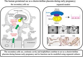
Abstract Image

人类妊娠囊作为绒毛膜胎盘在妊娠早期的作用;次级卵黄囊和类器官模型。
卵黄囊在系统发育上是胚胎外膜中最古老的,在许多物种的早期妊娠过程中,卵黄囊在营养传递方面发挥着重要作用。在人类,这一功能在很大程度上被认为是残余的,部分原因是次级卵黄囊从未与绒毛膜囊的内表面接触过。相反,它被充满液体的胚外联合体与绒毛膜分离,并通过相对较长的卵黄管与发育中的胚胎相连。然而,胚腔液中含有丰富的营养物质和关键辅助因子,包括叶酸和抗氧化剂,这些物质来自母体血浆和子宫内膜腺体。最近的大量测序发现,这些配体中存在大量转运蛋白的编码转录本。越来越多的证据表明,人类的次级卵黄囊在器官形成的关键阶段发挥着转运组织营养的关键作用,同时也在转运医疗药物和可替宁等化学物质方面发挥着关键作用。因此,我们提出,早期胎盘绒毛、腹腔和卵黄囊在妊娠最初几周结合起来发挥绒毛胎盘的生理功能。我们从小鼠卵黄囊中提取了有机体作为模型系统的原理验证,该模型系统可用于回答人类卵黄囊在妊娠头三个月作为母胎交换界面的功能能力方面的许多问题。
本文章由计算机程序翻译,如有差异,请以英文原文为准。
求助全文
约1分钟内获得全文 求助全文
来源期刊
Developmental biology
Developmental biology 生物-发育生物学
CiteScore
5.30
自引率
3.70%
发文量
182
审稿时长
1.5 months
期刊介绍: Developmental Biology (DB) publishes original research on mechanisms of development, differentiation, and growth in animals and plants at the molecular, cellular, genetic and evolutionary levels. Areas of particular emphasis include transcriptional control mechanisms, embryonic patterning, cell-cell interactions, growth factors and signal transduction, and regulatory hierarchies in developing plants and animals.
×
引用
GB/T 7714-2015
复制
MLA
复制
APA
复制
导出至
BibTeX EndNote RefMan NoteFirst NoteExpress
×
提示
您的信息不完整,为了账户安全,请先补充。
现在去补充
×
提示
您因"违规操作"
具体请查看互助需知
我知道了
×
提示
确定
请完成安全验证×
copy
已复制链接
快去分享给好友吧!
我知道了
右上角分享
点击右上角分享
0
联系我们:info@booksci.cn Book学术提供免费学术资源搜索服务,方便国内外学者检索中英文文献。致力于提供最便捷和优质的服务体验。 Copyright © 2023 布克学术 All rights reserved.
京ICP备2023020795号-1
ghs 京公网安备 11010802042870号
Book学术文献互助
Book学术文献互助群
群 号:481959085
Book学术官方微信