Leukocyte Platelet-Rich Plasma-Derived Exosomes Restrained Macrophages Viability and Induced Apoptosis, NO Generation, and M1 Polarization

IF 3.1 4区 医学 Q3 IMMUNOLOGY
Xiong Li, Feifei Guo, Jiehua Deng, Jiasong Li, Jie Zhang, Ming Fu, Hui Fan
{"title":"Leukocyte Platelet-Rich Plasma-Derived Exosomes Restrained Macrophages Viability and Induced Apoptosis, NO Generation, and M1 Polarization","authors":"Xiong Li,&nbsp;Feifei Guo,&nbsp;Jiehua Deng,&nbsp;Jiasong Li,&nbsp;Jie Zhang,&nbsp;Ming Fu,&nbsp;Hui Fan","doi":"10.1002/iid3.70064","DOIUrl":null,"url":null,"abstract":"<div>\n \n \n <section>\n \n <h3> Background</h3>\n \n <p>Chronic refractory wounds refer to wounds that cannot be repaired timely. Platelet-rich plasma (PRP) has significant potential in chronic wound healing therapy. The exosomes isolated from PRP were proved to exhibit more effectiveness than PRP. However, the therapeutic potential of exosomes from PRP on chronic refractory wounds remained elusive. Hence, this study aimed to clarify the action of exosomes from PRP on chronic refractory wounds by evaluating the response of macrophages to exosomes.</p>\n </section>\n \n <section>\n \n <h3> Methods</h3>\n \n <p>Pure platelet-rich plasma (P-PRP) and leukocyte platelet-rich plasma (L-PRP) were prepared from the fasting venous blood of healthy volunteers. Exosomes were extracted from P-PRP and L-PRP using ultracentrifugation and identified by transmission electron microscopy (TEM), nanoparticle tracking analysis (NTA), and western blot. Macrophages were obtained by inducing THP-1 cells with phorbol-12-myristate-13 acetate (PMA). The internalization of exosomes into macrophages was observed utilizing confocal laser scanning microscopy after being labeled with PKH67. Cell viability was determined by CCK-8 assay. Cell apoptosis was measured utilizing a flow cytometer. The polarization status of M1 and M2 macrophages were evaluated by detecting their markers. Nitric oxide (NO) detection was conducted using the commercial kit.</p>\n </section>\n \n <section>\n \n <h3> Results</h3>\n \n <p>Exosomes from P-PRP and L-PRP were absorbed by macrophages. Exosomes from L-PRP restrained viability and induced apoptosis of macrophages. Besides, exosomes from P-PRP promoted M2 polarization, and exosomes from L-PRP promoted M1 polarization. Furthermore, exosomes from L-PRP promoted NO generation of macrophages.</p>\n </section>\n \n <section>\n \n <h3> Conclusion</h3>\n \n <p>Exosomes from L-PRP restrained viability, induced apoptosis and NO generation of macrophages, and promoted M1 polarization, while exosomes from P-PRP increased M2 polarization. The exosomes from L-PRP presented a more effective effect on macrophages than that from P-PRP, making it a promising strategy for chronic refractory wound management.</p>\n </section>\n </div>","PeriodicalId":13289,"journal":{"name":"Immunity, Inflammation and Disease","volume":"12 11","pages":""},"PeriodicalIF":3.1000,"publicationDate":"2024-11-15","publicationTypes":"Journal Article","fieldsOfStudy":null,"isOpenAccess":false,"openAccessPdf":"https://www.ncbi.nlm.nih.gov/pmc/articles/PMC11565605/pdf/","citationCount":"0","resultStr":null,"platform":"Semanticscholar","paperid":null,"PeriodicalName":"Immunity, Inflammation and Disease","FirstCategoryId":"3","ListUrlMain":"https://onlinelibrary.wiley.com/doi/10.1002/iid3.70064","RegionNum":4,"RegionCategory":"医学","ArticlePicture":[],"TitleCN":null,"AbstractTextCN":null,"PMCID":null,"EPubDate":"","PubModel":"","JCR":"Q3","JCRName":"IMMUNOLOGY","Score":null,"Total":0}
引用次数: 0

Abstract

Background

Chronic refractory wounds refer to wounds that cannot be repaired timely. Platelet-rich plasma (PRP) has significant potential in chronic wound healing therapy. The exosomes isolated from PRP were proved to exhibit more effectiveness than PRP. However, the therapeutic potential of exosomes from PRP on chronic refractory wounds remained elusive. Hence, this study aimed to clarify the action of exosomes from PRP on chronic refractory wounds by evaluating the response of macrophages to exosomes.

Methods

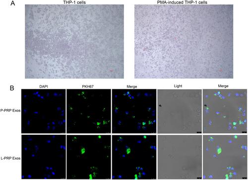
Pure platelet-rich plasma (P-PRP) and leukocyte platelet-rich plasma (L-PRP) were prepared from the fasting venous blood of healthy volunteers. Exosomes were extracted from P-PRP and L-PRP using ultracentrifugation and identified by transmission electron microscopy (TEM), nanoparticle tracking analysis (NTA), and western blot. Macrophages were obtained by inducing THP-1 cells with phorbol-12-myristate-13 acetate (PMA). The internalization of exosomes into macrophages was observed utilizing confocal laser scanning microscopy after being labeled with PKH67. Cell viability was determined by CCK-8 assay. Cell apoptosis was measured utilizing a flow cytometer. The polarization status of M1 and M2 macrophages were evaluated by detecting their markers. Nitric oxide (NO) detection was conducted using the commercial kit.

Results

Exosomes from P-PRP and L-PRP were absorbed by macrophages. Exosomes from L-PRP restrained viability and induced apoptosis of macrophages. Besides, exosomes from P-PRP promoted M2 polarization, and exosomes from L-PRP promoted M1 polarization. Furthermore, exosomes from L-PRP promoted NO generation of macrophages.

Conclusion

Exosomes from L-PRP restrained viability, induced apoptosis and NO generation of macrophages, and promoted M1 polarization, while exosomes from P-PRP increased M2 polarization. The exosomes from L-PRP presented a more effective effect on macrophages than that from P-PRP, making it a promising strategy for chronic refractory wound management.
Abstract Image

白细胞富血小板血浆衍生的外泌体抑制巨噬细胞活力并诱导凋亡、NO生成和M1极化
背景:慢性难治性伤口是指无法及时修复的伤口。富血小板血浆(PRP)在慢性伤口愈合治疗中具有巨大潜力。事实证明,从血小板丰富血浆中分离出的外泌体比血小板丰富血浆更有效。然而,从 PRP 中分离出的外泌体对慢性难治性伤口的治疗潜力仍然难以捉摸。因此,本研究旨在通过评估巨噬细胞对外泌体的反应,阐明从血小板丰富血浆中提取的外泌体对慢性难治性伤口的作用:方法:从健康志愿者的空腹静脉血中制备富含血小板的纯血浆(P-PRP)和富含白细胞的血小板血浆(L-PRP)。使用超速离心法从P-PRP和L-PRP中提取外泌体,并通过透射电子显微镜(TEM)、纳米颗粒追踪分析(NTA)和Western印迹进行鉴定。用光滑醇-12-肉豆蔻酸-13 乙酸酯(PMA)诱导 THP-1 细胞获得巨噬细胞。用 PKH67 标记外泌体后,利用激光扫描共聚焦显微镜观察外泌体在巨噬细胞中的内化情况。细胞活力通过 CCK-8 试验测定。细胞凋亡用流式细胞仪测定。通过检测 M1 和 M2 巨噬细胞的标记物来评估它们的极化状态。使用商业试剂盒检测一氧化氮(NO):结果:P-PRP和L-PRP的外泌体被巨噬细胞吸收。L-PRP 的外泌体抑制巨噬细胞的活力并诱导其凋亡。此外,P-PRP 的外泌体可促进 M2 极化,L-PRP 的外泌体可促进 M1 极化。此外,L-PRP的外泌体还能促进巨噬细胞产生NO:结论:L-PRP的外泌体抑制了巨噬细胞的活力,诱导了巨噬细胞的凋亡和NO生成,并促进了M1极化,而P-PRP的外泌体增加了M2极化。L-PRP的外泌体对巨噬细胞的作用比P-PRP的外泌体更有效,因此L-PRP有望成为慢性难治性伤口治疗的一种策略。
本文章由计算机程序翻译,如有差异,请以英文原文为准。
求助全文
约1分钟内获得全文 求助全文
来源期刊
Immunity, Inflammation and Disease
Immunity, Inflammation and Disease Medicine-Immunology and Allergy
CiteScore
3.60
自引率
0.00%
发文量
146
审稿时长
8 weeks
期刊介绍: Immunity, Inflammation and Disease is a peer-reviewed, open access, interdisciplinary journal providing rapid publication of research across the broad field of immunology. Immunity, Inflammation and Disease gives rapid consideration to papers in all areas of clinical and basic research. The journal is indexed in Medline and the Science Citation Index Expanded (part of Web of Science), among others. It welcomes original work that enhances the understanding of immunology in areas including: • cellular and molecular immunology • clinical immunology • allergy • immunochemistry • immunogenetics • immune signalling • immune development • imaging • mathematical modelling • autoimmunity • transplantation immunology • cancer immunology
×
引用
GB/T 7714-2015
复制
MLA
复制
APA
复制
导出至
BibTeX EndNote RefMan NoteFirst NoteExpress
×
提示
您的信息不完整,为了账户安全,请先补充。
现在去补充
×
提示
您因"违规操作"
具体请查看互助需知
我知道了
×
提示
确定
请完成安全验证×
copy
已复制链接
快去分享给好友吧!
我知道了
右上角分享
点击右上角分享
0
联系我们:info@booksci.cn Book学术提供免费学术资源搜索服务,方便国内外学者检索中英文文献。致力于提供最便捷和优质的服务体验。 Copyright © 2023 布克学术 All rights reserved.
京ICP备2023020795号-1
ghs 京公网安备 11010802042870号
Book学术文献互助
Book学术文献互助群
群 号:481959085
Book学术官方微信