Age-dependent cerebral vasodilation induced by volatile anesthetics is mediated by NG2+ vascular mural cells

IF 5.2 1区 生物学 Q1 BIOLOGY
Hang Zhou, Viola Neudecker, Jose F. Perez-Zoghbi, Ansgar M. Brambrink, Guang Yang
{"title":"Age-dependent cerebral vasodilation induced by volatile anesthetics is mediated by NG2+ vascular mural cells","authors":"Hang Zhou, Viola Neudecker, Jose F. Perez-Zoghbi, Ansgar M. Brambrink, Guang Yang","doi":"10.1038/s42003-024-07200-7","DOIUrl":null,"url":null,"abstract":"Anesthesia can influence cerebral blood flow by altering vessel diameter. Using in vivo two-photon imaging, we examined the effects of volatile anesthetics, sevoflurane and isoflurane, on vessel diameter in young and adult mice. Our results show that these anesthetics induce robust dilation of cortical arterioles and arteriole-proximate capillaries in adult mice, with milder effects in juveniles and no dilation in infants. This anesthesia-induced vasodilation correlates with decreased cytosolic Ca2+ levels in NG2+ vascular mural cells. Optogenetic manipulation of these cells bidirectionally regulates vessel diameter, and their ablation abolishes the vasodilatory response to anesthetics. In immature brains, NG2+ mural cells are fewer in number and express lower levels of Kir6.1, a subunit of ATP-sensitive potassium channels. This likely contributes to the age-dependent differences in vasodilation, as Kir6.1 activation promotes, while its inhibition reduces, anesthesia-induced vasodilation. These findings highlight the essential role of NG2+ mural cells in mediating anesthesia-induced cerebral vasodilation. Live animal imaging reveals age-dependent cerebral vasodilatory responses to volatile anesthetics, pronounced in adult mice and diminished or absent in developing brains. These effects are mediated by NG2+ mural cells and Kir6.1 signaling.","PeriodicalId":10552,"journal":{"name":"Communications Biology","volume":" ","pages":"1-16"},"PeriodicalIF":5.2000,"publicationDate":"2024-11-15","publicationTypes":"Journal Article","fieldsOfStudy":null,"isOpenAccess":false,"openAccessPdf":"https://www.ncbi.nlm.nih.gov/pmc/articles/PMC11568297/pdf/","citationCount":"0","resultStr":null,"platform":"Semanticscholar","paperid":null,"PeriodicalName":"Communications Biology","FirstCategoryId":"99","ListUrlMain":"https://www.nature.com/articles/s42003-024-07200-7","RegionNum":1,"RegionCategory":"生物学","ArticlePicture":[],"TitleCN":null,"AbstractTextCN":null,"PMCID":null,"EPubDate":"","PubModel":"","JCR":"Q1","JCRName":"BIOLOGY","Score":null,"Total":0}
引用次数: 0

Abstract

Anesthesia can influence cerebral blood flow by altering vessel diameter. Using in vivo two-photon imaging, we examined the effects of volatile anesthetics, sevoflurane and isoflurane, on vessel diameter in young and adult mice. Our results show that these anesthetics induce robust dilation of cortical arterioles and arteriole-proximate capillaries in adult mice, with milder effects in juveniles and no dilation in infants. This anesthesia-induced vasodilation correlates with decreased cytosolic Ca2+ levels in NG2+ vascular mural cells. Optogenetic manipulation of these cells bidirectionally regulates vessel diameter, and their ablation abolishes the vasodilatory response to anesthetics. In immature brains, NG2+ mural cells are fewer in number and express lower levels of Kir6.1, a subunit of ATP-sensitive potassium channels. This likely contributes to the age-dependent differences in vasodilation, as Kir6.1 activation promotes, while its inhibition reduces, anesthesia-induced vasodilation. These findings highlight the essential role of NG2+ mural cells in mediating anesthesia-induced cerebral vasodilation. Live animal imaging reveals age-dependent cerebral vasodilatory responses to volatile anesthetics, pronounced in adult mice and diminished or absent in developing brains. These effects are mediated by NG2+ mural cells and Kir6.1 signaling.
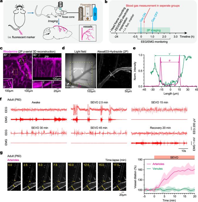
Abstract Image

挥发性麻醉剂诱导的年龄依赖性脑血管扩张是由 NG2+ 血管壁细胞介导的。
麻醉可通过改变血管直径来影响脑血流量。我们利用体内双光子成像技术研究了挥发性麻醉剂七氟醚和异氟醚对幼鼠和成年小鼠血管直径的影响。我们的研究结果表明,这些麻醉剂可诱导成年小鼠皮层动脉和动脉近端毛细血管的强烈扩张,对幼鼠的影响较小,而对婴儿则没有扩张作用。这种麻醉诱导的血管扩张与 NG2+ 血管壁细胞的细胞质 Ca2+ 水平下降有关。对这些细胞进行光遗传学操作可双向调节血管直径,而对它们的消减则可消除对麻醉剂的血管扩张反应。在未成熟的大脑中,NG2+ 壁细胞数量较少,表达的 ATP 敏感钾通道亚基 Kir6.1 水平较低。这可能是造成血管扩张的年龄依赖性差异的原因,因为Kir6.1的激活会促进麻醉诱导的血管扩张,而抑制则会减少这种扩张。这些发现凸显了 NG2+ 壁细胞在介导麻醉诱导的脑血管扩张中的重要作用。
本文章由计算机程序翻译,如有差异,请以英文原文为准。
求助全文
约1分钟内获得全文 求助全文
来源期刊
Communications Biology
Communications Biology Medicine-Medicine (miscellaneous)
CiteScore
8.60
自引率
1.70%
发文量
1233
审稿时长
13 weeks
期刊介绍: Communications Biology is an open access journal from Nature Research publishing high-quality research, reviews and commentary in all areas of the biological sciences. Research papers published by the journal represent significant advances bringing new biological insight to a specialized area of research.
×
引用
GB/T 7714-2015
复制
MLA
复制
APA
复制
导出至
BibTeX EndNote RefMan NoteFirst NoteExpress
×
提示
您的信息不完整,为了账户安全,请先补充。
现在去补充
×
提示
您因"违规操作"
具体请查看互助需知
我知道了
×
提示
确定
请完成安全验证×
copy
已复制链接
快去分享给好友吧!
我知道了
右上角分享
点击右上角分享
0
联系我们:info@booksci.cn Book学术提供免费学术资源搜索服务,方便国内外学者检索中英文文献。致力于提供最便捷和优质的服务体验。 Copyright © 2023 布克学术 All rights reserved.
京ICP备2023020795号-1
ghs 京公网安备 11010802042870号
Book学术文献互助
Book学术文献互助群
群 号:481959085
Book学术官方微信