Jemin Hwang, Beung Chul Ahn, So Hyeon Ji, Ho-Shin Gwak
{"title":"Complete Remission of Dural-Based Leptomeningeal Metastasis in Patient With Non-Small Cell Lung Cancer by Osimertinib.","authors":"Jemin Hwang, Beung Chul Ahn, So Hyeon Ji, Ho-Shin Gwak","doi":"10.14791/btrt.2024.0034","DOIUrl":null,"url":null,"abstract":"<p><p>We report complete remission of dural-based leptomeningeal metastasis (LM) in an 80-year-old female patient with non-small cell lung cancer (NSCLC) by osimertinib. She was diagnosed with NSCLC (adenocarcinoma, T4N3M1a) 8 years ago. Mutation analysis of biopsied tissue revealed exon 19 deletion positive, and gefitinib was prescribed. Follow-up chest CT showed a radiological response, and whole-body positron emission tomography 3 years later revealed the disappearance of the previous high-uptake lesions. The medication was continued for maintenance but stopped 4 years later due to intolerable dermatitis. Two years after discontinuing chemotherapy, the patient had a gait disturbance, and brain MRI revealed a right cerebellar mass (diameter [d]=3 cm) with peritumoral edema, compatible with solitary brain metastasis. Retromastoid suboccipital craniotomy and gross total removal of the dura-attached lesion were performed. As the systemic cancer status evaluation revealed no radiological cancer lesion, only tumor bed radiation therapy was given (4,000 cGy/10 fractions) without re-introducing gefitinib. She was followed with a brain MRI at 6-month intervals, and a brain MRI 2 years postoperatively revealed a dural-based extra-axial mass in the left prepontine cistern (d=2.2 cm). Serial cerebrospinal fluid (CSF) cytology was positive for cancer cells. Upon LM diagnosis, the third-generation receptor tyrosine kinase inhibitor osimertinib was given. Two-month follow-up CSF cytology and five consecutive tests over 14 months demonstrated negative conversion. Five-month follow-up brain MRI revealed near complete remission of dural-based LM, and the response was maintained until the 13-month follow-up brain MRI.</p>","PeriodicalId":72453,"journal":{"name":"Brain tumor research and treatment","volume":"12 4","pages":"245-249"},"PeriodicalIF":0.0000,"publicationDate":"2024-10-01","publicationTypes":"Journal Article","fieldsOfStudy":null,"isOpenAccess":false,"openAccessPdf":"https://www.ncbi.nlm.nih.gov/pmc/articles/PMC11570083/pdf/","citationCount":"0","resultStr":null,"platform":"Semanticscholar","paperid":null,"PeriodicalName":"Brain tumor research and treatment","FirstCategoryId":"1085","ListUrlMain":"https://doi.org/10.14791/btrt.2024.0034","RegionNum":0,"RegionCategory":null,"ArticlePicture":[],"TitleCN":null,"AbstractTextCN":null,"PMCID":null,"EPubDate":"","PubModel":"","JCR":"","JCRName":"","Score":null,"Total":0}
引用次数: 0
Abstract
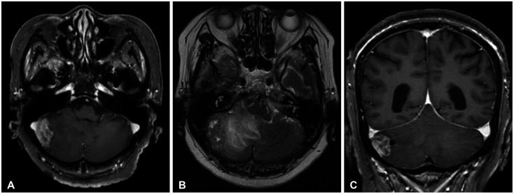
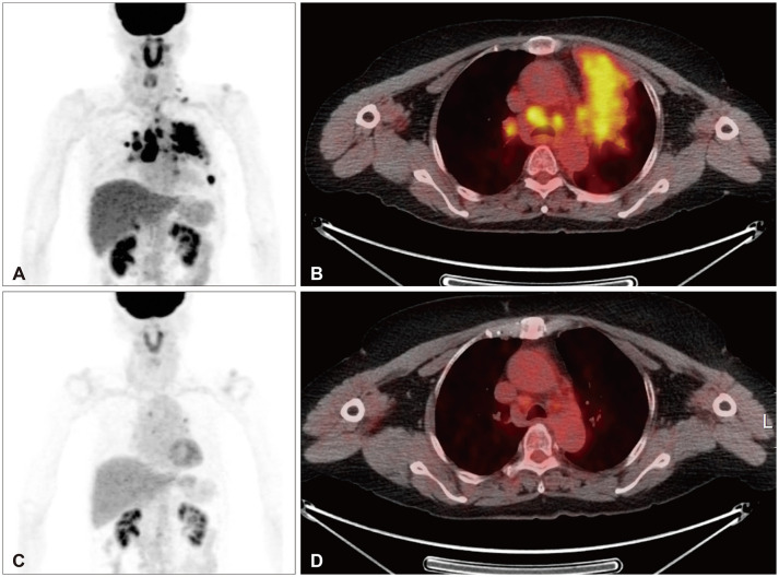
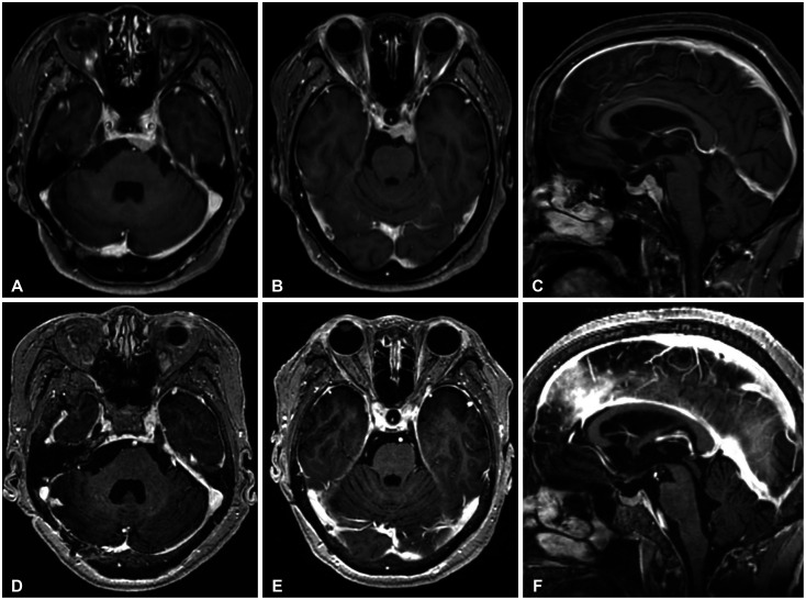
We report complete remission of dural-based leptomeningeal metastasis (LM) in an 80-year-old female patient with non-small cell lung cancer (NSCLC) by osimertinib. She was diagnosed with NSCLC (adenocarcinoma, T4N3M1a) 8 years ago. Mutation analysis of biopsied tissue revealed exon 19 deletion positive, and gefitinib was prescribed. Follow-up chest CT showed a radiological response, and whole-body positron emission tomography 3 years later revealed the disappearance of the previous high-uptake lesions. The medication was continued for maintenance but stopped 4 years later due to intolerable dermatitis. Two years after discontinuing chemotherapy, the patient had a gait disturbance, and brain MRI revealed a right cerebellar mass (diameter [d]=3 cm) with peritumoral edema, compatible with solitary brain metastasis. Retromastoid suboccipital craniotomy and gross total removal of the dura-attached lesion were performed. As the systemic cancer status evaluation revealed no radiological cancer lesion, only tumor bed radiation therapy was given (4,000 cGy/10 fractions) without re-introducing gefitinib. She was followed with a brain MRI at 6-month intervals, and a brain MRI 2 years postoperatively revealed a dural-based extra-axial mass in the left prepontine cistern (d=2.2 cm). Serial cerebrospinal fluid (CSF) cytology was positive for cancer cells. Upon LM diagnosis, the third-generation receptor tyrosine kinase inhibitor osimertinib was given. Two-month follow-up CSF cytology and five consecutive tests over 14 months demonstrated negative conversion. Five-month follow-up brain MRI revealed near complete remission of dural-based LM, and the response was maintained until the 13-month follow-up brain MRI.

 求助内容:
求助内容: 应助结果提醒方式:


