Anatomical Significance of the Patent Foramen Ovale by Real-Time 3D TEE in Cryptogenic Stroke and Migraine

IF 1.6 4区 医学 Q3 CARDIAC & CARDIOVASCULAR SYSTEMS
Li Wang, Haibo Sun, Han Shen
{"title":"Anatomical Significance of the Patent Foramen Ovale by Real-Time 3D TEE in Cryptogenic Stroke and Migraine","authors":"Li Wang,&nbsp;Haibo Sun,&nbsp;Han Shen","doi":"10.1111/echo.70018","DOIUrl":null,"url":null,"abstract":"<div>\n \n \n <section>\n \n <h3> Background</h3>\n \n <p>The transesophageal echocardiogram (TEE) is the standard imaging modality for confirming the presence or absence of patent foramen ovale. PFO is a flap valve depending on the pressure change between the left and right atrium, which can help determine whether to open. 3D-TEE was shown to optimize the visualization of PFO. There is a causal association between PFO and unexplained stroke. It seems that 3D-TEE can present a high-risk PFO morphological feature, which seems to show more than just being easier to open.</p>\n </section>\n \n <section>\n \n <h3> Methods</h3>\n \n <p>In total, 134 consecutive patients with cryptogenic stroke or migraine who had suspected PFO and underwent c-TCD, TTE, and c-TEE were included in this study. TEE confirmed the PFO. The right-to-left shunt (RLS) grade of PFO at rest and abdominal compression Valsalva maneuver was detected by c-TEE.</p>\n </section>\n \n <section>\n \n <h3> Results</h3>\n \n <p>The long diameter of FO (1.74 ± 0.3 vs. 1.60 ± 0.4, <i>p</i> = 0.039), the short diameter of FO (1.12 ± 0.3 vs. 1.00 ± 0.3, <i>p</i> = 0.036), perimeter of FO (4.62 ± 0.7 vs. 4.22 ± 1.0, <i>p</i> = 0.026), and area (1.80 ± 0.8 vs. 1.35 ± 0.8, <i>p</i> = 0.05) of the FO were significantly larger in the larger RLS group. In group of CS, a larger proportion of Eustachian valve or a Chiari's network (14.3% vs. 3.5%, <i>p</i> = 0.036), a larger proportion of in the left funnelform (55.1% vs. 16.3%, <i>p</i> &lt; 0.001), a longer length of the PFO tunnel (13.4 ± 4.4 vs. 7.8 ± 2.5, <i>p</i> &lt; 0.001), a lower IVC-PFO angle (16.4 ± 3.4 vs. 20.3 ± 7.7, <i>p</i> = 0.001), a higher proportion of LA multiple exits of the tunnel (46.9% vs. 14.3%, <i>p</i> &lt; 0.001). Multivariate regression analysis showed that male gender (HR: 4.026, 95% CI: 0.883–18.361, <i>p</i> = 0.072), age (HR: 1.076, 95% CI: 1.002–1.155, <i>p</i> = 0.045), the left funnelform (HR: 7.299, 95% CI: 1.585–33.618, <i>p</i> = 0.011), a longer length of the PFO tunnel (HR: 1.843, 95% CI: 1.404–2.418, <i>p</i> &lt; 0.001) and multiple exits of the tunnel of LA (HR: 8.544, 95% CI: 1.595–45.754, <i>p</i> = 0.012) increased the risk of cerebral infarction. The cut-off value calculated by ROC for the diagnosis of high-risk PFO was that the length of the PFO tunnel was 12 mm and the left funnelform combined with multiple exits of the left atrial (sensitivity was 92%, specificity was 90%). The area under the curve of the combined index versus PoPE score (0.932 vs. 0.736) relative to the RoPE score was statistically significant.</p>\n </section>\n \n <section>\n \n <h3> Conclusions</h3>\n \n <p>TEE has shown outstanding advantages in displaying the specific morphological characteristics of PFO. The left funnelform, a longer length of the PFO tunnel, and multiple exits of the tunnel of LA are associated with an increasing risk of CS in anatomical PFO respect.</p>\n </section>\n </div>","PeriodicalId":50558,"journal":{"name":"Echocardiography-A Journal of Cardiovascular Ultrasound and Allied Techniques","volume":"41 11","pages":""},"PeriodicalIF":1.6000,"publicationDate":"2024-11-13","publicationTypes":"Journal Article","fieldsOfStudy":null,"isOpenAccess":false,"openAccessPdf":"https://onlinelibrary.wiley.com/doi/epdf/10.1111/echo.70018","citationCount":"0","resultStr":null,"platform":"Semanticscholar","paperid":null,"PeriodicalName":"Echocardiography-A Journal of Cardiovascular Ultrasound and Allied Techniques","FirstCategoryId":"3","ListUrlMain":"https://onlinelibrary.wiley.com/doi/10.1111/echo.70018","RegionNum":4,"RegionCategory":"医学","ArticlePicture":[],"TitleCN":null,"AbstractTextCN":null,"PMCID":null,"EPubDate":"","PubModel":"","JCR":"Q3","JCRName":"CARDIAC & CARDIOVASCULAR SYSTEMS","Score":null,"Total":0}
引用次数: 0

Abstract

Background

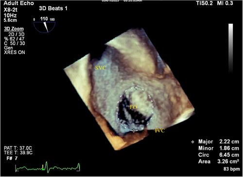
The transesophageal echocardiogram (TEE) is the standard imaging modality for confirming the presence or absence of patent foramen ovale. PFO is a flap valve depending on the pressure change between the left and right atrium, which can help determine whether to open. 3D-TEE was shown to optimize the visualization of PFO. There is a causal association between PFO and unexplained stroke. It seems that 3D-TEE can present a high-risk PFO morphological feature, which seems to show more than just being easier to open.

Methods

In total, 134 consecutive patients with cryptogenic stroke or migraine who had suspected PFO and underwent c-TCD, TTE, and c-TEE were included in this study. TEE confirmed the PFO. The right-to-left shunt (RLS) grade of PFO at rest and abdominal compression Valsalva maneuver was detected by c-TEE.

Results

The long diameter of FO (1.74 ± 0.3 vs. 1.60 ± 0.4, p = 0.039), the short diameter of FO (1.12 ± 0.3 vs. 1.00 ± 0.3, p = 0.036), perimeter of FO (4.62 ± 0.7 vs. 4.22 ± 1.0, p = 0.026), and area (1.80 ± 0.8 vs. 1.35 ± 0.8, p = 0.05) of the FO were significantly larger in the larger RLS group. In group of CS, a larger proportion of Eustachian valve or a Chiari's network (14.3% vs. 3.5%, p = 0.036), a larger proportion of in the left funnelform (55.1% vs. 16.3%, p < 0.001), a longer length of the PFO tunnel (13.4 ± 4.4 vs. 7.8 ± 2.5, p < 0.001), a lower IVC-PFO angle (16.4 ± 3.4 vs. 20.3 ± 7.7, p = 0.001), a higher proportion of LA multiple exits of the tunnel (46.9% vs. 14.3%, p < 0.001). Multivariate regression analysis showed that male gender (HR: 4.026, 95% CI: 0.883–18.361, p = 0.072), age (HR: 1.076, 95% CI: 1.002–1.155, p = 0.045), the left funnelform (HR: 7.299, 95% CI: 1.585–33.618, p = 0.011), a longer length of the PFO tunnel (HR: 1.843, 95% CI: 1.404–2.418, p < 0.001) and multiple exits of the tunnel of LA (HR: 8.544, 95% CI: 1.595–45.754, p = 0.012) increased the risk of cerebral infarction. The cut-off value calculated by ROC for the diagnosis of high-risk PFO was that the length of the PFO tunnel was 12 mm and the left funnelform combined with multiple exits of the left atrial (sensitivity was 92%, specificity was 90%). The area under the curve of the combined index versus PoPE score (0.932 vs. 0.736) relative to the RoPE score was statistically significant.

Conclusions

TEE has shown outstanding advantages in displaying the specific morphological characteristics of PFO. The left funnelform, a longer length of the PFO tunnel, and multiple exits of the tunnel of LA are associated with an increasing risk of CS in anatomical PFO respect.
Abstract Image

通过实时三维 TEE 观察隐源性中风和偏头痛中闭孔卵巢的解剖学意义。
背景:经食道超声心动图(TEE)是确认是否存在卵圆孔未闭的标准成像方式。PFO 是一个瓣膜,取决于左心房和右心房之间的压力变化,这有助于确定是否开放。三维超声心动图可优化 PFO 的可视化。PFO 与不明原因中风之间存在因果关系。3D-TEE 似乎可以呈现高风险 PFO 的形态特征,这似乎不仅仅是显示更容易打开:本研究共纳入了 134 名疑似 PFO 的隐源性卒中或偏头痛患者,他们都接受了 c-TCD、TTE 和 c-TEE。TEE 证实了 PFO。c-TEE 检测了 PFO 在静息和腹部压迫 Valsalva 动作时的右向左分流(RLS)等级:结果:FO 长径(1.74 ± 0.3 vs. 1.60 ± 0.4,p = 0.039)、FO 短径(1.12 ± 0.3 vs. 1.00 ± 0.3,p = 0.036)、FO 周径(4.62 ± 0.7 vs. 4.22 ± 1.0,p = 0.026)和面积(1.80 ± 0.8 vs. 1.35 ± 0.8,p = 0.05)均明显大于较大的 RLS 组。在 CS 组中,咽鼓管瓣或奇氏网络的比例较大(14.3% vs. 3.5%,P = 0.036),左侧漏斗状体的比例较大(55.1% vs. 16.3%,P < 0.001),PFO 通道的长度较长(13.4 ± 4.4 vs. 7.8 ± 2.5,p < 0.001),IVC-PFO角度较小(16.4 ± 3.4 vs. 20.3 ± 7.7,p = 0.001),隧道LA多个出口的比例较高(46.9% vs. 14.3%,p < 0.001)。多变量回归分析显示,男性(HR:4.026,95% CI:0.883-18.361,p = 0.072)、年龄(HR:1.076,95% CI:1.002-1.155,p = 0.045)、左侧漏斗状(HR:7.299,95% CI:1.585-33.618,p = 0.011)、PFO 通道长度较长(HR:1.843,95% CI:1.404-2.418,P<0.001)和 LA 通道有多个出口(HR:8.544,95% CI:1.595-45.754,P=0.012)会增加脑梗死的风险。通过 ROC 计算得出的诊断高危 PFO 的临界值是 PFO 通道长度为 12 mm,左心房漏斗形合并左心房多出口(敏感性为 92%,特异性为 90%)。综合指数与 PoPE 评分(0.932 对 0.736)相对于 RoPE 评分的曲线下面积具有统计学意义:结论:TEE 在显示 PFO 的具体形态特征方面具有突出优势。左侧漏斗形、较长的 PFO 通道和 LA 通道的多个出口与解剖型 PFO 的 CS 风险增加有关。
本文章由计算机程序翻译,如有差异,请以英文原文为准。
求助全文
约1分钟内获得全文 求助全文
来源期刊
CiteScore
2.40
自引率
6.70%
发文量
211
审稿时长
3-6 weeks
期刊介绍: Echocardiography: A Journal of Cardiovascular Ultrasound and Allied Techniques is the official publication of the International Society of Cardiovascular Ultrasound. Widely recognized for its comprehensive peer-reviewed articles, case studies, original research, and reviews by international authors. Echocardiography keeps its readership of echocardiographers, ultrasound specialists, and cardiologists well informed of the latest developments in the field.
×
引用
GB/T 7714-2015
复制
MLA
复制
APA
复制
导出至
BibTeX EndNote RefMan NoteFirst NoteExpress
×
提示
您的信息不完整,为了账户安全,请先补充。
现在去补充
×
提示
您因"违规操作"
具体请查看互助需知
我知道了
×
提示
确定
请完成安全验证×
copy
已复制链接
快去分享给好友吧!
我知道了
右上角分享
点击右上角分享
0
联系我们:info@booksci.cn Book学术提供免费学术资源搜索服务,方便国内外学者检索中英文文献。致力于提供最便捷和优质的服务体验。 Copyright © 2023 布克学术 All rights reserved.
京ICP备2023020795号-1
ghs 京公网安备 11010802042870号
Book学术文献互助
Book学术文献互助群
群 号:481959085
Book学术官方微信