Teresa Cristina de Castro Ramos Sarmet Dos Santos, Mai-Lan Ho, Maria de Fatima Vasco Aragão, Renata Artimos de Oliveira Vianna, Alexandre Ribeiro Fernandes, Alair Augusto Sarmet Moreira Damas Dos Santos, Claudete Aparecida Araújo Cardoso
{"title":"Brain MRI in infants exposed to the Zika virus, with one-year follow-up: expanding the phenotype.","authors":"Teresa Cristina de Castro Ramos Sarmet Dos Santos, Mai-Lan Ho, Maria de Fatima Vasco Aragão, Renata Artimos de Oliveira Vianna, Alexandre Ribeiro Fernandes, Alair Augusto Sarmet Moreira Damas Dos Santos, Claudete Aparecida Araújo Cardoso","doi":"10.1590/0100-3984.2024.0014","DOIUrl":null,"url":null,"abstract":"<p><strong>Objective: </strong>To analyze longitudinal changes between two brain magnetic resonance imaging (MRI) exams performed one year apart in symptomatic infants with congenital Zika syndrome (CZS) and normocephalic infants exposed to the Zika virus (ZIKV) prenatally.</p><p><strong>Materials and methods: </strong>This was a prospective observational study. Infants born to women who tested positive for ZIKV on reverse transcription-quantitative polymerase chain reaction during pregnancy were classified into two groups: symptomatic infants with CZS and asymptomatic infants. All of the infants underwent brain MRI at presentation and after one year of follow-up. All MRI scans were evaluated independently by a pediatric radiologist and a pediatric neuroradiologist, and the infants underwent clinical monitoring by a pediatric neurologist.</p><p><strong>Results: </strong>The sample included 36 infants exposed to ZIKV perinatally. Therefore, a total of 72 MRI scans were evaluated. Among the 36 infants included a diagnosis of CZS was made in 25 (69.4%), of whom 18 presented with a combination of classic findings (including reduced brain volume, subcortical calcifications, brainstem hypoplasia, malformations of the corpus callosum, malformations of cortical development, and ventriculomegaly), as well as atypical findings such as hyperintense foci in the white matter on T2-weighted sequences. Of those same 25 infants, seven presented with mild lesions. Of the 11 normocephalic patients, five (13.9%) had atypical findings such as hyperintense foci in the white matter on T2-weighted sequences and no other manifestations of CZS, although there was mild neurological involvement. Six (16.6%) of the 36 patients had completely normal MRI scans with no neurological changes. No disease progression was observed during follow-up.</p><p><strong>Conclusion: </strong>In infants exposed to ZIKV perinatally, the frequency of classic and atypical findings on brain MRI seems to be associated with the neurological status. Brain MRI is an important diagnostic tool in the evaluation and monitoring of patients with congenital infection, because intracranial changes other than microcephaly can occur.</p>","PeriodicalId":20842,"journal":{"name":"Radiologia Brasileira","volume":"57 ","pages":"e20240014"},"PeriodicalIF":0.0000,"publicationDate":"2024-11-07","publicationTypes":"Journal Article","fieldsOfStudy":null,"isOpenAccess":false,"openAccessPdf":"https://www.ncbi.nlm.nih.gov/pmc/articles/PMC11559958/pdf/","citationCount":"0","resultStr":null,"platform":"Semanticscholar","paperid":null,"PeriodicalName":"Radiologia Brasileira","FirstCategoryId":"1085","ListUrlMain":"https://doi.org/10.1590/0100-3984.2024.0014","RegionNum":0,"RegionCategory":null,"ArticlePicture":[],"TitleCN":null,"AbstractTextCN":null,"PMCID":null,"EPubDate":"2024/1/1 0:00:00","PubModel":"eCollection","JCR":"Q3","JCRName":"Medicine","Score":null,"Total":0}
引用次数: 0
Abstract
Objective: To analyze longitudinal changes between two brain magnetic resonance imaging (MRI) exams performed one year apart in symptomatic infants with congenital Zika syndrome (CZS) and normocephalic infants exposed to the Zika virus (ZIKV) prenatally.
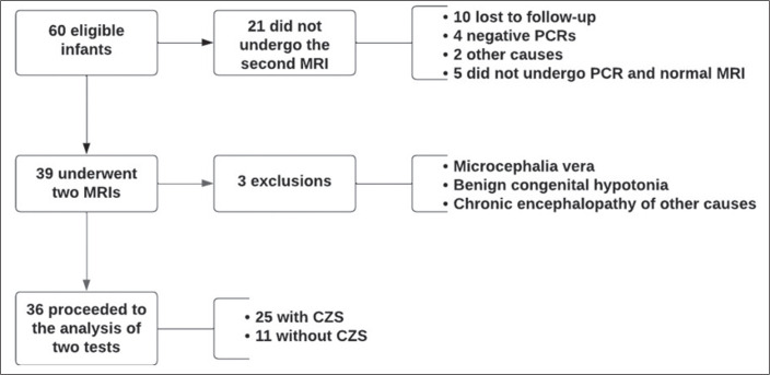
Materials and methods: This was a prospective observational study. Infants born to women who tested positive for ZIKV on reverse transcription-quantitative polymerase chain reaction during pregnancy were classified into two groups: symptomatic infants with CZS and asymptomatic infants. All of the infants underwent brain MRI at presentation and after one year of follow-up. All MRI scans were evaluated independently by a pediatric radiologist and a pediatric neuroradiologist, and the infants underwent clinical monitoring by a pediatric neurologist.
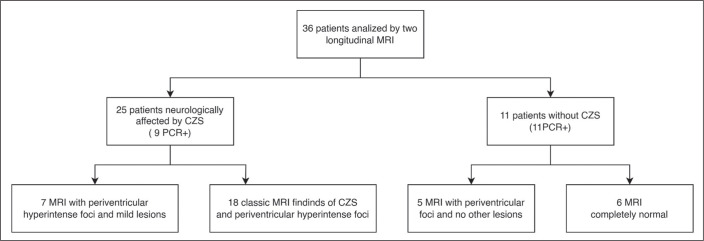
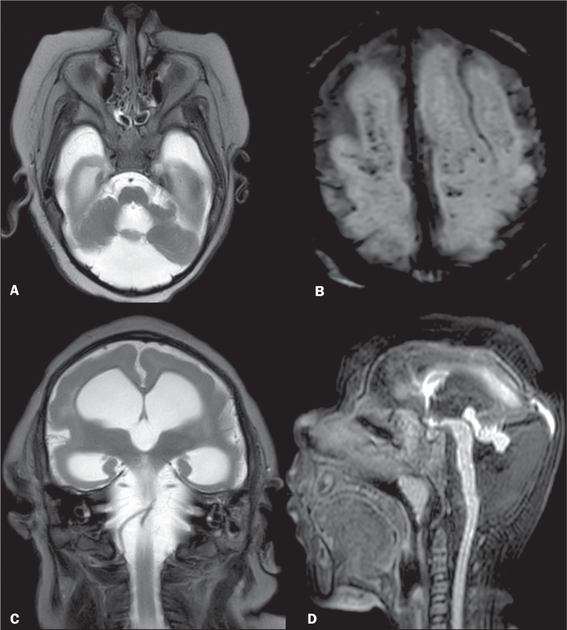
Results: The sample included 36 infants exposed to ZIKV perinatally. Therefore, a total of 72 MRI scans were evaluated. Among the 36 infants included a diagnosis of CZS was made in 25 (69.4%), of whom 18 presented with a combination of classic findings (including reduced brain volume, subcortical calcifications, brainstem hypoplasia, malformations of the corpus callosum, malformations of cortical development, and ventriculomegaly), as well as atypical findings such as hyperintense foci in the white matter on T2-weighted sequences. Of those same 25 infants, seven presented with mild lesions. Of the 11 normocephalic patients, five (13.9%) had atypical findings such as hyperintense foci in the white matter on T2-weighted sequences and no other manifestations of CZS, although there was mild neurological involvement. Six (16.6%) of the 36 patients had completely normal MRI scans with no neurological changes. No disease progression was observed during follow-up.
Conclusion: In infants exposed to ZIKV perinatally, the frequency of classic and atypical findings on brain MRI seems to be associated with the neurological status. Brain MRI is an important diagnostic tool in the evaluation and monitoring of patients with congenital infection, because intracranial changes other than microcephaly can occur.

 求助内容:
求助内容: 应助结果提醒方式:


