Machine learning identifies arrhythmogenic features of QRS fragmentation in human cardiomyopathy: Implications for improving risk stratification
IF 5.7
2区 医学
Q1 CARDIAC & CARDIOVASCULAR SYSTEMS
引用次数: 0
Abstract
Background
Heterogeneous ventricular activation can provide the substrate for ventricular arrhythmias (VA), but its manifestation on the electrocardiogram (ECG) as a risk stratifier is not well-defined.
Objective
To characterize the spatiotemporal features of QRS peaks that best predict VA in patients with cardiomyopathy (CM) using machine learning (ML).
Methods
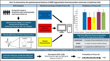
Prospectively enrolled CM patients with prophylactic defibrillators (n=95) underwent digital, high-resolution ECG recordings during intrinsic rhythm and ventricular pacing at 100 to 120 beats/min. Intra QRS peaks in the signal-averaged precordial leads were identified and their characteristics (amplitude, width, and timing within the QRS) were transformed into 4-bin histograms. Random forest models of these characteristics in each lead alongside clinical data were trained on 76 patients and tested on 19 patients with cross-validation to determine the features that predicted VA.
Results
Patients were followed up for 45 (22–48) months, and 21% had VA. The individual machine learning (ML) models determined (area under the receiver operating characteristic curve [AUROC]) intrinsic QRS peak amplitude (0.88), width (0.78), and location (0.84) to all predict VA. In a combined model including all QRS peak characteristics, peaks with amplitude < 31 μV in V1, a width of 4 to 8 ms in V1, and location in the final quarter of the QRS of V1 were most predictive. Neither clinical data nor QRS peak characteristics assessed during ventricular pacing improved VA prediction when combined with intrinsic QRS peak characteristics.
Conclusions
Arrhythmogenic QRS fragmentation is characterized by narrow, low-amplitude peaks in the terminal QRS of lead V1. These features alone without clinical variables or ventricular pacing are sufficient to accurately risk stratify CM patients.

机器学习识别人类心肌病 QRS 分段的致心律失常特征:对改进风险分层的意义。
背景:异质性心室激活可为室性心律失常(VA)提供基质,但其在心电图上作为风险分层指标的表现尚未明确:目的:利用机器学习(ML)描述最能预测心肌病(CM)患者室性心律失常的 QRS 峰时空特征:前瞻性入组的使用预防性除颤器的心肌病患者(95 人)在固有节律和 100-120bpm 的心室起搏期间接受了数字高分辨率心电图记录。在信号平均的心前导联中识别出 QRS 内峰,并将其特征(QRS 内的振幅、宽度和时间)转换成 4 分段直方图。在 76 名患者身上训练了每个导联中这些特征的随机森林模型以及临床数据,并在 19 名患者身上进行了交叉验证测试,以确定预测 VA 的特征:结果:对患者进行了 45(22-48)个月的随访,21% 的患者获得了 VA。单个 ML 模型确定(AUROC)固有 QRS 峰值振幅(0.88)、宽度(0.78)和位置(0.84)均可预测 VA。在一个包括所有 QRS 峰特征的组合模型中,振幅峰得出了结论:致心律失常性 QRS 分裂的特征是 V1 导联 QRS 末端出现狭窄、低振幅的峰值。仅凭这些特征而不考虑临床变量或心室起搏,就足以对 CM 患者进行准确的风险分层。
本文章由计算机程序翻译,如有差异,请以英文原文为准。
求助全文
约1分钟内获得全文
求助全文
来源期刊

Heart rhythm
医学-心血管系统
CiteScore
10.50
自引率
5.50%
发文量
1465
审稿时长
24 days
期刊介绍:
HeartRhythm, the official Journal of the Heart Rhythm Society and the Cardiac Electrophysiology Society, is a unique journal for fundamental discovery and clinical applicability.
HeartRhythm integrates the entire cardiac electrophysiology (EP) community from basic and clinical academic researchers, private practitioners, engineers, allied professionals, industry, and trainees, all of whom are vital and interdependent members of our EP community.
The Heart Rhythm Society is the international leader in science, education, and advocacy for cardiac arrhythmia professionals and patients, and the primary information resource on heart rhythm disorders. Its mission is to improve the care of patients by promoting research, education, and optimal health care policies and standards.
 求助内容:
求助内容: 应助结果提醒方式:


