Odontogenic carcinosarcoma of the mandible: A case report

IF 0.6 Q3 MEDICINE, GENERAL & INTERNAL
Pouyan Aminishakib, Farzaneh Mosavat, Mahsa Bayati, Ata Garajei
{"title":"Odontogenic carcinosarcoma of the mandible: A case report","authors":"Pouyan Aminishakib,&nbsp;Farzaneh Mosavat,&nbsp;Mahsa Bayati,&nbsp;Ata Garajei","doi":"10.1002/ccr3.9554","DOIUrl":null,"url":null,"abstract":"<div>\n \n <section>\n \n <h3> Key Clinical Message</h3>\n \n <p>Odontogenic carcinosarcoma, a rare and challenging diagnosis, was identified in a 60-year-old male through histopathology, revealing a biphasic neoplasm with malignant epithelial and mesenchymal components. Surgical resection is crucial for management, highlighting the importance of vigilant postoperative follow-up to ensure early detection of any recurrence.</p>\n </section>\n \n <section>\n \n \n <p>One rare mixed malignant odontogenic tumor is odontogenic carcinosarcoma, which comprises malignant epithelial and mesenchymal components. Diagnosing odontogenic carcinosarcoma is challenging due to its rarity and atypical clinical presentation. This study reports a 60-year-old male patient who presented with a painless swelling on the right side of his face and experienced facial asymmetry for 6 months, ultimately diagnosed with odontogenic carcinosarcoma. A biphasic neoplasm with malignant alterations in both epithelial and mesenchymal components was identified upon histopathological examination. MRI imaging showed an expansile multilobulated lytic lesion with cortical erosion and extraosseous extension in the posterior region of the right mandibular body and ramus. Following contrast administration, homogeneous lesion enhancement was observed, with a few small non-enhancing necrotic areas in central parts. The patient subsequently underwent a right hemi-mandibulectomy with resection of adjacent soft tissues and neck dissection due to lymph node involvement. The resulting defect was reconstructed using a pectoralis major flap. No recurrence or metastasis was reported during the 6-month follow-up, reinforcing the positive results. This case highlights the importance of recognizing odontogenic carcinosarcoma and underscores the challenges in diagnosing and managing this rare tumor. Early identification and aggressive treatment can lead to positive outcomes, as evidenced by the absence of recurrence or metastasis in this patient during the follow-up period.</p>\n </section>\n </div>","PeriodicalId":10327,"journal":{"name":"Clinical Case Reports","volume":"12 11","pages":""},"PeriodicalIF":0.6000,"publicationDate":"2024-11-11","publicationTypes":"Journal Article","fieldsOfStudy":null,"isOpenAccess":false,"openAccessPdf":"https://www.ncbi.nlm.nih.gov/pmc/articles/PMC11554428/pdf/","citationCount":"0","resultStr":null,"platform":"Semanticscholar","paperid":null,"PeriodicalName":"Clinical Case Reports","FirstCategoryId":"1085","ListUrlMain":"https://onlinelibrary.wiley.com/doi/10.1002/ccr3.9554","RegionNum":0,"RegionCategory":null,"ArticlePicture":[],"TitleCN":null,"AbstractTextCN":null,"PMCID":null,"EPubDate":"","PubModel":"","JCR":"Q3","JCRName":"MEDICINE, GENERAL & INTERNAL","Score":null,"Total":0}
引用次数: 0

Abstract

Key Clinical Message

Odontogenic carcinosarcoma, a rare and challenging diagnosis, was identified in a 60-year-old male through histopathology, revealing a biphasic neoplasm with malignant epithelial and mesenchymal components. Surgical resection is crucial for management, highlighting the importance of vigilant postoperative follow-up to ensure early detection of any recurrence.

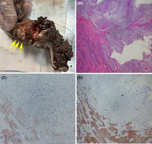
One rare mixed malignant odontogenic tumor is odontogenic carcinosarcoma, which comprises malignant epithelial and mesenchymal components. Diagnosing odontogenic carcinosarcoma is challenging due to its rarity and atypical clinical presentation. This study reports a 60-year-old male patient who presented with a painless swelling on the right side of his face and experienced facial asymmetry for 6 months, ultimately diagnosed with odontogenic carcinosarcoma. A biphasic neoplasm with malignant alterations in both epithelial and mesenchymal components was identified upon histopathological examination. MRI imaging showed an expansile multilobulated lytic lesion with cortical erosion and extraosseous extension in the posterior region of the right mandibular body and ramus. Following contrast administration, homogeneous lesion enhancement was observed, with a few small non-enhancing necrotic areas in central parts. The patient subsequently underwent a right hemi-mandibulectomy with resection of adjacent soft tissues and neck dissection due to lymph node involvement. The resulting defect was reconstructed using a pectoralis major flap. No recurrence or metastasis was reported during the 6-month follow-up, reinforcing the positive results. This case highlights the importance of recognizing odontogenic carcinosarcoma and underscores the challenges in diagnosing and managing this rare tumor. Early identification and aggressive treatment can lead to positive outcomes, as evidenced by the absence of recurrence or metastasis in this patient during the follow-up period.
Abstract Image

下颌骨牙源性癌肉瘤:病例报告
关键临床信息:一名60岁男性的牙源性癌肉瘤是一种罕见且具有挑战性的诊断,通过组织病理学检查发现,该肿瘤是一种具有恶性上皮和间质成分的双相肿瘤。手术切除是治疗的关键,强调术后密切随访的重要性,以确保及早发现复发。摘要:牙源性癌肉瘤是一种罕见的混合性恶性牙源性肿瘤,由恶性上皮和间质成分组成。由于其罕见性和不典型的临床表现,诊断牙源性癌肉瘤具有挑战性。本研究报告了一名60岁的男性患者,他因右侧面部无痛性肿胀而就诊,面部不对称症状持续了6个月,最终被诊断为牙源性癌肉瘤。组织病理学检查发现,这是一种双相肿瘤,上皮和间质均有恶性改变。核磁共振成像显示,右下颌骨体和臼齿后部出现扩张性多叶溶解性病变,伴有皮质侵蚀和骨外扩展。使用造影剂后,病变呈均匀强化,中央部位有几个小的非强化坏死区。由于淋巴结受累,患者随后接受了右半下颌骨切除术,并切除了邻近软组织和颈部清扫术。使用胸大肌皮瓣对缺损部位进行了重建。在 6 个月的随访期间,未发现复发或转移,巩固了积极的治疗效果。该病例强调了识别牙源性癌肉瘤的重要性,并突出了诊断和治疗这种罕见肿瘤所面临的挑战。正如该患者在随访期间没有复发或转移所证明的那样,早期识别和积极治疗可带来积极的结果。
本文章由计算机程序翻译,如有差异,请以英文原文为准。
求助全文
约1分钟内获得全文 求助全文
来源期刊
Clinical Case Reports
Clinical Case Reports MEDICINE, GENERAL & INTERNAL-
自引率
14.30%
发文量
1268
审稿时长
13 weeks
期刊介绍: Clinical Case Reports is different from other case report journals. Our aim is to directly improve global health and increase clinical understanding using case reports to convey important best practice information. We welcome case reports from all areas of Medicine, Nursing, Dentistry, and Veterinary Science and may include: -Any clinical case or procedure which illustrates an important best practice teaching message -Any clinical case or procedure which illustrates the appropriate use of an important clinical guideline or systematic review. As well as: -The management of novel or very uncommon diseases -A common disease presenting in an uncommon way -An uncommon disease masquerading as something more common -Cases which expand understanding of disease pathogenesis -Cases where the teaching point is based on an error -Cases which allow us to re-think established medical lore -Unreported adverse effects of interventions (drug, procedural, or other).
×
引用
GB/T 7714-2015
复制
MLA
复制
APA
复制
导出至
BibTeX EndNote RefMan NoteFirst NoteExpress
×
提示
您的信息不完整,为了账户安全,请先补充。
现在去补充
×
提示
您因"违规操作"
具体请查看互助需知
我知道了
×
提示
确定
请完成安全验证×
copy
已复制链接
快去分享给好友吧!
我知道了
右上角分享
点击右上角分享
0
联系我们:info@booksci.cn Book学术提供免费学术资源搜索服务,方便国内外学者检索中英文文献。致力于提供最便捷和优质的服务体验。 Copyright © 2023 布克学术 All rights reserved.
京ICP备2023020795号-1
ghs 京公网安备 11010802042870号
Book学术文献互助
Book学术文献互助群
群 号:481959085
Book学术官方微信