High intensity interval training as a therapy: Mitophagy restoration in breast cancer
IF 3.8
3区 生物学
Q2 BIOCHEMISTRY & MOLECULAR BIOLOGY
引用次数: 0
Abstract
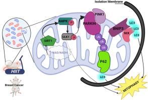
Recent studies have highlighted the role of mitophagy in tumorigenesis. This study aimed to investigate the effects of high-intensity interval training (HIIT) on mitophagy in tumor tissues of mice with breast cancer. Twenty-eight female BALB/c mice were randomly assigned to four groups: Healthy Control (CO), Cancer (CA), Exercise (EX), and Cancer + Exercise (CA + EX). Mammary tumors were induced in the CA and CA + EX groups via 4T1 cell injections. Upon confirmation of tumor formation, the EX and CA + EX groups underwent 8 weeks (40 sessions) of HIIT, comprising 4–10 intervals of running at 80–100 % of maximum speed. The expression levels of mitophagy-related proteins, including parkin, PTEN-induced putative kinase 1 (PINK1), NIP3-like protein X (NIX), BCL2 interacting protein-3 (BINP3), microtubule-associated protein light chain 3-I (LC3-I), microtubule-associated protein light chain 3-II (LC3-II), AMP-activated protein kinase (AMPK), Unc-51 like autophagy activating kinase-1 (ULK1), and sirtuin-1 (SIRT1), were measured in breast and tumor tissues. Tumor volume relative to body weight was assessed weekly during the eight-week HIIT intervention. Protein expression of parkin, PINK1, NIX, BINP3, LC3-II, LC3-I, AMPK, ULK1, and SIRT1 was reduced in the breast tissue of the CA group, while HIIT restored expression levels across all measured variables (P < 0.01). Additionally, tumor volume relative to body weight was significantly lower in the CA + EX group compared to the CA group from weeks 3–8 (P < 0.01). These findings suggest that breast cancer suppresses mitophagy, yet HIIT effectively reverses this suppression, potentially reducing tumor burden. HIIT may thus represent a promising therapeutic strategy for managing breast cancer.

将高强度间歇训练作为一种疗法:恢复乳腺癌患者的丝裂噬功能
最近的研究强调了有丝分裂在肿瘤发生中的作用。本研究旨在探讨高强度间歇训练(HIIT)对乳腺癌小鼠肿瘤组织中有丝分裂的影响。28只雌性BALB/c小鼠被随机分为四组:健康对照组(CO)、癌症组(CA)、运动组(EX)和癌症+运动组(CA + EX)。CA 组和 CA + EX 组通过注射 4T1 细胞诱发乳腺肿瘤。确认肿瘤形成后,EX组和CA + EX组进行为期8周(40次)的HIIT训练,包括4-10次以80-100%的最大速度跑步。有丝分裂相关蛋白的表达水平,包括parkin、PTEN诱导的假定激酶1(PINK1)、NIP3样蛋白X(NIX)、BCL2相互作用蛋白-3(BINP3)、微管相关蛋白轻链3-I(LC3-I)、乳腺和肿瘤组织中的微管相关蛋白轻链 3-II (LC3-II)、AMP 激活蛋白激酶 (AMPK)、Unc-51 类自噬激活激酶-1 (ULK1) 和 sirtuin-1 (SIRT1)。在为期八周的 HIIT 干预期间,每周评估肿瘤体积相对于体重的情况。在 CA 组的乳腺组织中,parkin、PINK1、NIX、BINP3、LC3-II、LC3-I、AMPK、ULK1 和 SIRT1 的蛋白表达量减少,而 HIIT 恢复了所有测量变量的表达水平(P
本文章由计算机程序翻译,如有差异,请以英文原文为准。
求助全文
约1分钟内获得全文
求助全文
来源期刊

Archives of biochemistry and biophysics
生物-生化与分子生物学
CiteScore
7.40
自引率
0.00%
发文量
245
审稿时长
26 days
期刊介绍:
Archives of Biochemistry and Biophysics publishes quality original articles and reviews in the developing areas of biochemistry and biophysics.
Research Areas Include:
• Enzyme and protein structure, function, regulation. Folding, turnover, and post-translational processing
• Biological oxidations, free radical reactions, redox signaling, oxygenases, P450 reactions
• Signal transduction, receptors, membrane transport, intracellular signals. Cellular and integrated metabolism.
 求助内容:
求助内容: 应助结果提醒方式:


