Is There a Correlation Between the Location of the Clot in the Pulmonary Arterial Tree with the Location of the Mucus Plug in the Pulmonary Bronchial Tree in Patients with chronic obstructive pulmonary disease Experiencing Pulmonary Embolism? Novel Findings.
{"title":"Is There a Correlation Between the Location of the Clot in the Pulmonary Arterial Tree with the Location of the Mucus Plug in the Pulmonary Bronchial Tree in Patients with chronic obstructive pulmonary disease Experiencing Pulmonary Embolism? Novel Findings.","authors":"Neda Akhoundi, Alireza Siami, Zahra Naseri, Mahlagha Amirbakhtiarvand","doi":"10.5152/ThoracResPract.2024.23137","DOIUrl":null,"url":null,"abstract":"<p><strong>Objective: </strong>The study aimed to investigate the impact of mucus plugs on the localization of clots in pulmonary embolisms among chronic obstructive pulmonary disease (COPD) patients.</p><p><strong>Material and methods: </strong>The retrospective study examined 200 participants diagnosed with both COPD and pulmonary embolism. Of these, 100 patients exhibited mucus plugs in the segmental and subsegmental branches of the pulmonary bronchial tree, while the remaining 100 did not, using computed tomography images for diagnosis. Data collection encompassed a comprehensive review of patient records, including medical history and imaging reports, to determine the presence of mucus plugs and the localization of clots in pulmonary embolism cases.</p><p><strong>Results: </strong>Patients with mucus plugs exhibited a notably longer duration of COPD (P = .021) and a higher mean pulmonary arterial occlusion index (23 vs. 12, P = .001). Moreover, the prevalence of clots in major pulmonary arteries was significantly elevated in the mucus plug group compared to the non-mucus plug group (P < .05). Conversely, patients without mucus plugs displayed a higher incidence of clots in segmental and subsegmental arteries (P < .001). Strong positive correlations existed between mucus plugs in segmental branches and clots in major pulmonary arteries, with moderate to strong correlation coefficients (0.51 to 0.62, P < .05). Additionally, strong negative correlations were observed between mucus plugs in segmental branches and clots in segmental and subsegmental arteries, with correlation coefficients (CC) ranging from -0.74 to -0.76 (P < .05). Similarly, a significant negative correlation was noted between mucus plugs in subsegmental branches and clots in subsegmental arteries (CC: -0.68 and -0.71, P < .05).</p><p><strong>Conclusion: </strong>The results suggest that mucus plugs may be associated with increased severity of COPD, higher pulmonary arterial occlusion index, and altered clot distribution within the pulmonary artery tree. These findings emphasize the importance of recognizing mucus plugs as a key consideration in COPD assessment and management, potentially influencing disease severity, vascular remodeling, and thrombotic risk management.</p>","PeriodicalId":75221,"journal":{"name":"Thoracic research and practice","volume":"25 6","pages":"203-208"},"PeriodicalIF":0.6000,"publicationDate":"2024-11-01","publicationTypes":"Journal Article","fieldsOfStudy":null,"isOpenAccess":false,"openAccessPdf":"https://www.ncbi.nlm.nih.gov/pmc/articles/PMC11565401/pdf/","citationCount":"0","resultStr":null,"platform":"Semanticscholar","paperid":null,"PeriodicalName":"Thoracic research and practice","FirstCategoryId":"1085","ListUrlMain":"https://doi.org/10.5152/ThoracResPract.2024.23137","RegionNum":0,"RegionCategory":null,"ArticlePicture":[],"TitleCN":null,"AbstractTextCN":null,"PMCID":null,"EPubDate":"","PubModel":"","JCR":"0","JCRName":"RESPIRATORY SYSTEM","Score":null,"Total":0}
引用次数: 0
Abstract
Objective: The study aimed to investigate the impact of mucus plugs on the localization of clots in pulmonary embolisms among chronic obstructive pulmonary disease (COPD) patients.
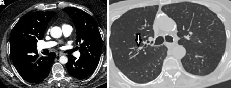
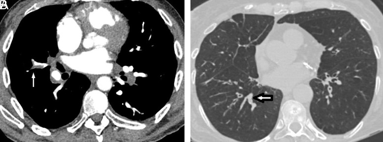
Material and methods: The retrospective study examined 200 participants diagnosed with both COPD and pulmonary embolism. Of these, 100 patients exhibited mucus plugs in the segmental and subsegmental branches of the pulmonary bronchial tree, while the remaining 100 did not, using computed tomography images for diagnosis. Data collection encompassed a comprehensive review of patient records, including medical history and imaging reports, to determine the presence of mucus plugs and the localization of clots in pulmonary embolism cases.
Results: Patients with mucus plugs exhibited a notably longer duration of COPD (P = .021) and a higher mean pulmonary arterial occlusion index (23 vs. 12, P = .001). Moreover, the prevalence of clots in major pulmonary arteries was significantly elevated in the mucus plug group compared to the non-mucus plug group (P < .05). Conversely, patients without mucus plugs displayed a higher incidence of clots in segmental and subsegmental arteries (P < .001). Strong positive correlations existed between mucus plugs in segmental branches and clots in major pulmonary arteries, with moderate to strong correlation coefficients (0.51 to 0.62, P < .05). Additionally, strong negative correlations were observed between mucus plugs in segmental branches and clots in segmental and subsegmental arteries, with correlation coefficients (CC) ranging from -0.74 to -0.76 (P < .05). Similarly, a significant negative correlation was noted between mucus plugs in subsegmental branches and clots in subsegmental arteries (CC: -0.68 and -0.71, P < .05).
Conclusion: The results suggest that mucus plugs may be associated with increased severity of COPD, higher pulmonary arterial occlusion index, and altered clot distribution within the pulmonary artery tree. These findings emphasize the importance of recognizing mucus plugs as a key consideration in COPD assessment and management, potentially influencing disease severity, vascular remodeling, and thrombotic risk management.


 求助内容:
求助内容: 应助结果提醒方式:


