Posterior corneal elevation after small incision lenticule extraction (SMILE) in eyes of different myopia severity

IF 3.1 3区 医学 Q2 ONCOLOGY
Li Jiang , Changting Tang , Bao Shu , Ying Liu , Qingyan Zeng , Xiaohua Lei , Zhengwei Shen , Yijun Hu
{"title":"Posterior corneal elevation after small incision lenticule extraction (SMILE) in eyes of different myopia severity","authors":"Li Jiang ,&nbsp;Changting Tang ,&nbsp;Bao Shu ,&nbsp;Ying Liu ,&nbsp;Qingyan Zeng ,&nbsp;Xiaohua Lei ,&nbsp;Zhengwei Shen ,&nbsp;Yijun Hu","doi":"10.1016/j.pdpdt.2024.104392","DOIUrl":null,"url":null,"abstract":"<div><h3>Purpose</h3><div>To investigate the changes in posterior corneal elevation after small incision lenticule extraction (SMILE) surgery in eyes of different myopia severity.</div></div><div><h3>Methods</h3><div>A total of 141 eyes of 86 patients were recruited and followed at 1 month, 3 months, 6 months, and 12 months after SMILE surgery. The eyes were divided into four groups according to the spherical equivalent (SE): low myopia (LM, −3.00 D≤SE&lt;−0.50 D, 35 eyes), moderate myopia (MM, −6.00 D≤SE&lt;−3.00 D, 54 eyes) and high myopia (HM, −10.00 D≤SE&lt;−6.00 D, 52 eyes). Posterior corneal elevation was measured by Pentacam HR. Posterior corneal apex elevation (PCAE), posterior maximum elevation (PME), and posterior elevation at the thinnest point of the cornea (TPC) in the central 4-mm area were recorded and compared among different examination time points in the same group or among different groups at the same time point.</div></div><div><h3>Results</h3><div>Uncorrected distance visual acuity (UDVA) at 12 months was 20/20 or better in 100.00 % of eyes in LM group, 100.00 % in MM group and 96.15 % in HM group (<em>P</em> &gt; 0.05). There were significant reductions of post-operative PCAE compared to baseline in the LM group (<em>P</em> = 0.038) and HM group (<em>P</em>&lt;0.001), and the reduction was marginally significant in the MM group (<em>P</em> = 0.076). No statistically significant changes in PME were observed after SMILE surgery in the three groups (all <em>P</em> &gt; 0.05). TPC was significantly decreased after the surgery in the MM group (<em>P</em> = 0.022) and the HM group (<em>P</em>&lt;0.001), and was marginally significantly decreased in the LM group (<em>P</em> = 0.063). There was no significant difference in PCAE and PME among the three groups at baseline or any postoperative timepoint (all <em>P</em> &gt; 0.05). TPC was significantly different among the three groups at 3 months, 6 months and 12 months (all <em>P</em> &lt; 0.05), but not at baseline (<em>P</em> = 0.066) or 1 month (<em>P</em> = 0.080).</div></div><div><h3>Conclusion</h3><div>In the first year after SMILE surgery, PCAE was decreased in both LM and HM eyes, and TPC were decreased in MM and HM eyes, but PME was stable after the surgery.</div></div>","PeriodicalId":20141,"journal":{"name":"Photodiagnosis and Photodynamic Therapy","volume":"50 ","pages":"Article 104392"},"PeriodicalIF":3.1000,"publicationDate":"2024-11-04","publicationTypes":"Journal Article","fieldsOfStudy":null,"isOpenAccess":false,"openAccessPdf":"","citationCount":"0","resultStr":null,"platform":"Semanticscholar","paperid":null,"PeriodicalName":"Photodiagnosis and Photodynamic Therapy","FirstCategoryId":"3","ListUrlMain":"https://www.sciencedirect.com/science/article/pii/S1572100024004290","RegionNum":3,"RegionCategory":"医学","ArticlePicture":[],"TitleCN":null,"AbstractTextCN":null,"PMCID":null,"EPubDate":"","PubModel":"","JCR":"Q2","JCRName":"ONCOLOGY","Score":null,"Total":0}
引用次数: 0

Abstract

Purpose

To investigate the changes in posterior corneal elevation after small incision lenticule extraction (SMILE) surgery in eyes of different myopia severity.

Methods

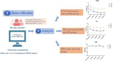
A total of 141 eyes of 86 patients were recruited and followed at 1 month, 3 months, 6 months, and 12 months after SMILE surgery. The eyes were divided into four groups according to the spherical equivalent (SE): low myopia (LM, −3.00 D≤SE<−0.50 D, 35 eyes), moderate myopia (MM, −6.00 D≤SE<−3.00 D, 54 eyes) and high myopia (HM, −10.00 D≤SE<−6.00 D, 52 eyes). Posterior corneal elevation was measured by Pentacam HR. Posterior corneal apex elevation (PCAE), posterior maximum elevation (PME), and posterior elevation at the thinnest point of the cornea (TPC) in the central 4-mm area were recorded and compared among different examination time points in the same group or among different groups at the same time point.

Results

Uncorrected distance visual acuity (UDVA) at 12 months was 20/20 or better in 100.00 % of eyes in LM group, 100.00 % in MM group and 96.15 % in HM group (P > 0.05). There were significant reductions of post-operative PCAE compared to baseline in the LM group (P = 0.038) and HM group (P<0.001), and the reduction was marginally significant in the MM group (P = 0.076). No statistically significant changes in PME were observed after SMILE surgery in the three groups (all P > 0.05). TPC was significantly decreased after the surgery in the MM group (P = 0.022) and the HM group (P<0.001), and was marginally significantly decreased in the LM group (P = 0.063). There was no significant difference in PCAE and PME among the three groups at baseline or any postoperative timepoint (all P > 0.05). TPC was significantly different among the three groups at 3 months, 6 months and 12 months (all P < 0.05), but not at baseline (P = 0.066) or 1 month (P = 0.080).

Conclusion

In the first year after SMILE surgery, PCAE was decreased in both LM and HM eyes, and TPC were decreased in MM and HM eyes, but PME was stable after the surgery.
Abstract Image

不同近视度数的眼睛在小切口人工晶体摘除术(SMILE)后角膜后部隆起的情况。
目的:研究不同近视度数的眼睛在接受小切口人工晶体摘除术(SMILE)后角膜后凸度的变化:方法:共招募了 86 名患者的 141 只眼睛,分别在 SMILE 手术后 1 个月、3 个月、6 个月和 12 个月进行随访。根据球面等效度数(SE)将患者分为四组:低度近视(LM,-3.00 D≤SEResults):12个月后,低度近视组、高度近视组和中度近视组的未矫正远视力(UDVA)分别达到或超过20/20的比例分别为100.00%、100.00%和96.15%(P>0.05)。LM 组(P=0.038)和 HM 组(P0.05)术后 PCAE 与基线相比明显下降。MM组(P=0.022)和HM组(P0.05)术后TPC明显下降。在 3 个月、6 个月和 12 个月时,三组的 TPC 有明显差异(均为 PC):SMILE手术后第一年,LM组和HM组的PCAE均下降,MM组和HM组的TPC均下降,但PME在术后保持稳定。
本文章由计算机程序翻译,如有差异,请以英文原文为准。
求助全文
约1分钟内获得全文 求助全文
来源期刊
CiteScore
5.80
自引率
24.20%
发文量
509
审稿时长
50 days
期刊介绍: Photodiagnosis and Photodynamic Therapy is an international journal for the dissemination of scientific knowledge and clinical developments of Photodiagnosis and Photodynamic Therapy in all medical specialties. The journal publishes original articles, review articles, case presentations, "how-to-do-it" articles, Letters to the Editor, short communications and relevant images with short descriptions. All submitted material is subject to a strict peer-review process.
×
引用
GB/T 7714-2015
复制
MLA
复制
APA
复制
导出至
BibTeX EndNote RefMan NoteFirst NoteExpress
×
提示
您的信息不完整,为了账户安全,请先补充。
现在去补充
×
提示
您因"违规操作"
具体请查看互助需知
我知道了
×
提示
确定
请完成安全验证×
copy
已复制链接
快去分享给好友吧!
我知道了
右上角分享
点击右上角分享
0
联系我们:info@booksci.cn Book学术提供免费学术资源搜索服务,方便国内外学者检索中英文文献。致力于提供最便捷和优质的服务体验。 Copyright © 2023 布克学术 All rights reserved.
京ICP备2023020795号-1
ghs 京公网安备 11010802042870号
Book学术文献互助
Book学术文献互助群
群 号:481959085
Book学术官方微信