Assessment of Pedicle screw-Rod implantation as an external fixation method for tibial osteotomy in a canine model

IF 1.9 Q2 AGRICULTURE, DAIRY & ANIMAL SCIENCE
{"title":"Assessment of Pedicle screw-Rod implantation as an external fixation method for tibial osteotomy in a canine model","authors":"","doi":"10.1016/j.vas.2024.100403","DOIUrl":null,"url":null,"abstract":"<div><div>Recent advancements in minimally invasive osteosynthesis have improved atraumatic techniques for bone fracture fixation. Pedicle screws are implants primarily used for the internal fixation of the spine. To our knowledge, no studies have assessed the use of Pedicle screw-Rod for fixing long bone fractures or osteotomies. Our study aimed to assess the efficiency and performance of this implant as an external fixation method for experimentally induced tibial fractures, offering a novel surgical approach to tibial fixation. With approval from the Institutional Animal Care Committee, eight healthy, intact male dogs weighing 20–22 kg and aged 10–12 months of mixed breeds underwent aseptic surgical fixation of tibial osteotomies with Pedicle screw-Rod configuration using a minimally invasive medial approach to the tibia. All dogs were placed in the single treatment group. Postoperative clinical and radiographic evaluations were performed. The fixation device functioned effectively until removal. Lameness was fully resolved in all animals by 21 days post-operation. Clinical union occurred at 5.80 ± 1.30 weeks, while complete bone union was achieved at 11.40 ± 1.51 weeks after surgery. Postoperative mechanical medial proximal and distal tibial angles were, 92.00° (92.00°, 91.50°) and 93.40° ± 1.14°, respectively. The tibial valgus was 5.20° ± 1.48°, and tibial plateau angles measured 22.00° (23.00°, 22.00°). There were no significant differences noted when comparing values before and after the operation. Postoperative rotational alignment was anatomical, with satisfactory bone apposition. The study found that using a Pedicle Screw-Rod configuration for non-articular tibial osteotomy fixation is effective without significant complications.</div></div>","PeriodicalId":37152,"journal":{"name":"Veterinary and Animal Science","volume":null,"pages":null},"PeriodicalIF":1.9000,"publicationDate":"2024-10-30","publicationTypes":"Journal Article","fieldsOfStudy":null,"isOpenAccess":false,"openAccessPdf":"","citationCount":"0","resultStr":null,"platform":"Semanticscholar","paperid":null,"PeriodicalName":"Veterinary and Animal Science","FirstCategoryId":"1085","ListUrlMain":"https://www.sciencedirect.com/science/article/pii/S2451943X2400070X","RegionNum":0,"RegionCategory":null,"ArticlePicture":[],"TitleCN":null,"AbstractTextCN":null,"PMCID":null,"EPubDate":"","PubModel":"","JCR":"Q2","JCRName":"AGRICULTURE, DAIRY & ANIMAL SCIENCE","Score":null,"Total":0}
引用次数: 0

Abstract

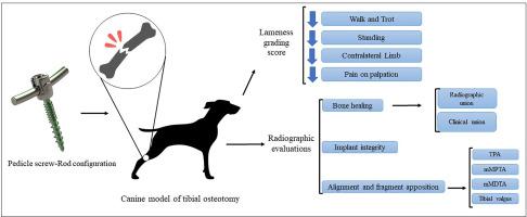
Recent advancements in minimally invasive osteosynthesis have improved atraumatic techniques for bone fracture fixation. Pedicle screws are implants primarily used for the internal fixation of the spine. To our knowledge, no studies have assessed the use of Pedicle screw-Rod for fixing long bone fractures or osteotomies. Our study aimed to assess the efficiency and performance of this implant as an external fixation method for experimentally induced tibial fractures, offering a novel surgical approach to tibial fixation. With approval from the Institutional Animal Care Committee, eight healthy, intact male dogs weighing 20–22 kg and aged 10–12 months of mixed breeds underwent aseptic surgical fixation of tibial osteotomies with Pedicle screw-Rod configuration using a minimally invasive medial approach to the tibia. All dogs were placed in the single treatment group. Postoperative clinical and radiographic evaluations were performed. The fixation device functioned effectively until removal. Lameness was fully resolved in all animals by 21 days post-operation. Clinical union occurred at 5.80 ± 1.30 weeks, while complete bone union was achieved at 11.40 ± 1.51 weeks after surgery. Postoperative mechanical medial proximal and distal tibial angles were, 92.00° (92.00°, 91.50°) and 93.40° ± 1.14°, respectively. The tibial valgus was 5.20° ± 1.48°, and tibial plateau angles measured 22.00° (23.00°, 22.00°). There were no significant differences noted when comparing values before and after the operation. Postoperative rotational alignment was anatomical, with satisfactory bone apposition. The study found that using a Pedicle Screw-Rod configuration for non-articular tibial osteotomy fixation is effective without significant complications.
Abstract Image

在犬模型中评估椎弓根螺钉-Rod 植入作为胫骨截骨外固定方法的效果
微创骨合成技术的最新进展改进了骨折固定的无创伤技术。椎弓根螺钉是主要用于脊柱内固定的植入物。据我们所知,还没有研究评估过椎弓根螺钉-Rod 用于固定长骨骨折或截骨。我们的研究旨在评估该植入物作为实验诱导的胫骨骨折外固定方法的效率和性能,为胫骨固定提供一种新的手术方法。经机构动物护理委员会批准,8 只体重 20-22 千克、年龄 10-12 个月的健康完好的混种雄性犬接受了无菌手术固定胫骨截骨,使用椎弓根螺钉-Rod 配置,采用微创胫骨内侧入路。所有犬只均为单一治疗组。术后进行了临床和放射学评估。固定装置在拆除前一直有效。术后 21 天,所有动物的跛行症状都完全消失。临床骨结合发生在术后 5.80 ± 1.30 周,完全骨结合发生在术后 11.40 ± 1.51 周。术后机械性胫骨内侧近端和远端角度分别为 92.00° (92.00°, 91.50°) 和 93.40° ± 1.14°。胫骨外翻角度为 5.20° ± 1.48°,胫骨平台角度为 22.00°(23.00°,22.00°)。手术前后的数值比较没有明显差异。术后的旋转对位符合解剖学原理,骨结合情况令人满意。研究发现,使用椎弓根螺钉-Rod配置进行非关节胫骨截骨固定效果显著,且无明显并发症。
本文章由计算机程序翻译,如有差异,请以英文原文为准。
求助全文
约1分钟内获得全文 求助全文
来源期刊
Veterinary and Animal Science
Veterinary and Animal Science Veterinary-Veterinary (all)
CiteScore
3.50
自引率
0.00%
发文量
43
审稿时长
47 days
×
引用
GB/T 7714-2015
复制
MLA
复制
APA
复制
导出至
BibTeX EndNote RefMan NoteFirst NoteExpress
×
提示
您的信息不完整,为了账户安全,请先补充。
现在去补充
×
提示
您因"违规操作"
具体请查看互助需知
我知道了
×
提示
确定
请完成安全验证×
copy
已复制链接
快去分享给好友吧!
我知道了
右上角分享
点击右上角分享
0
联系我们:info@booksci.cn Book学术提供免费学术资源搜索服务,方便国内外学者检索中英文文献。致力于提供最便捷和优质的服务体验。 Copyright © 2023 布克学术 All rights reserved.
京ICP备2023020795号-1
ghs 京公网安备 11010802042870号
Book学术文献互助
Book学术文献互助群
群 号:481959085
Book学术官方微信