In vivo performance of lean bioabsorbable Mg–Ca alloy X0 and comparison to WE43: Influence of surface modification and alloying content
IF 18
1区 医学
Q1 ENGINEERING, BIOMEDICAL
引用次数: 0
Abstract
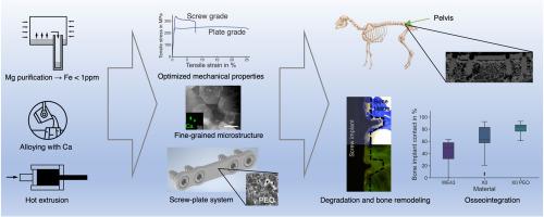
Magnesium alloys present a compelling prospect for absorbable implant materials in orthopedic and trauma surgery. This study evaluates an ultra-high purity, lean magnesium–calcium alloy (X0), both with and without plasma electrolytic oxidation (PEO) surface modification, in comparison to a clinically utilized WE43 magnesium alloy. It is shown that the mechanical properties of X0 can be tuned to yield a high-strength material suitable for bone screws (with an ultimate tensile strength of 336 MPa) or a ductile material appropriate for intraoperatively deformable plates (with an elongation at fracture of 24 %). Four plate-screw combinations were implanted onto the pelvic bones of six sheep without osteotomy for 8 weeks. Subsequent analysis utilized histology, micro-computed tomography, and light and electron microscopy. All implants exhibited signs of degradation and hydrogen-gas evolution, with PEO-coated X0 implants demonstrating the least volume loss and the most substantial new-bone formation on the implant surface and surrounding cancellous bone. Furthermore, the osteoconductive properties of the X0 implants, when uncoated, exceeded those of the uncoated WE43 implants, as evidenced by greater new-bone formation on the surface. This osteoconductivity was amplified with PEO surface modification, which mitigated gas evolution and enhanced osseointegration, encouraging bone apposition in the cancellous bone vicinity. These findings thus indicate that PEO-coated X0 implants hold substantial promise as a biocompatible and absorbable implant material.

瘦生物可吸收镁钙合金 X0 的体内性能以及与 WE43 的比较:表面改性和合金含量的影响
镁合金是整形外科和创伤外科中一种前景广阔的可吸收植入材料。本研究评估了一种超高纯度、贫镁钙合金(X0),将其与临床上使用的 WE43 镁合金进行了比较,前者经过和未经过等离子电解氧化(PEO)表面改性。结果表明,X0 的机械性能可以进行调整,以获得适合骨螺钉的高强度材料(极限抗拉强度为 336 兆帕)或适合术中可变形钢板的韧性材料(断裂伸长率为 24%)。将四种钢板-螺钉组合植入六只绵羊的骨盆骨中,不进行截骨手术,为期八周。随后利用组织学、微型计算机断层扫描以及光镜和电子显微镜进行了分析。所有植入物都出现了降解和氢气演化的迹象,其中 PEO 涂层 X0 植入物的体积损失最小,植入物表面和周围松质骨的新骨形成最多。此外,X0 植入体在未涂层时的骨传导性能超过了未涂层的 WE43 植入体,其表面形成的新骨更多就证明了这一点。这种骨传导性在 PEO 表面改性后得到增强,PEO 可减轻气体演化,增强骨结合,促进松质骨附近的骨附着。因此,这些研究结果表明,PEO 涂层 X0 植入物作为一种生物相容性和可吸收性植入材料具有广阔的前景。
本文章由计算机程序翻译,如有差异,请以英文原文为准。
求助全文
约1分钟内获得全文
求助全文
来源期刊

Bioactive Materials
Biochemistry, Genetics and Molecular Biology-Biotechnology
CiteScore
28.00
自引率
6.30%
发文量
436
审稿时长
20 days
期刊介绍:
Bioactive Materials is a peer-reviewed research publication that focuses on advancements in bioactive materials. The journal accepts research papers, reviews, and rapid communications in the field of next-generation biomaterials that interact with cells, tissues, and organs in various living organisms.
The primary goal of Bioactive Materials is to promote the science and engineering of biomaterials that exhibit adaptiveness to the biological environment. These materials are specifically designed to stimulate or direct appropriate cell and tissue responses or regulate interactions with microorganisms.
The journal covers a wide range of bioactive materials, including those that are engineered or designed in terms of their physical form (e.g. particulate, fiber), topology (e.g. porosity, surface roughness), or dimensions (ranging from macro to nano-scales). Contributions are sought from the following categories of bioactive materials:
Bioactive metals and alloys
Bioactive inorganics: ceramics, glasses, and carbon-based materials
Bioactive polymers and gels
Bioactive materials derived from natural sources
Bioactive composites
These materials find applications in human and veterinary medicine, such as implants, tissue engineering scaffolds, cell/drug/gene carriers, as well as imaging and sensing devices.
 求助内容:
求助内容: 应助结果提醒方式:


