Quantitative synthetic MRI for evaluation of hippocampus in patients with multiple sclerosis

IF 2.7 4区 医学 Q3 NEUROSCIENCES
Jing Huang , Yan Liang , Jiyuan Wang , Yi Shan , Cheng Zhao , Qiongge Li , Huiqing Dong , Jie Lu
{"title":"Quantitative synthetic MRI for evaluation of hippocampus in patients with multiple sclerosis","authors":"Jing Huang ,&nbsp;Yan Liang ,&nbsp;Jiyuan Wang ,&nbsp;Yi Shan ,&nbsp;Cheng Zhao ,&nbsp;Qiongge Li ,&nbsp;Huiqing Dong ,&nbsp;Jie Lu","doi":"10.1016/j.brainres.2024.149298","DOIUrl":null,"url":null,"abstract":"<div><h3>Objective</h3><div>To identify early changes in hippocampal quantitative parameters in multiple sclerosis (MS) patients using synthetic MRI, and to correlate these changes with clinical variables.</div></div><div><h3>Methods</h3><div>45 MS patients and 26 healthy controls (HCs) underwent synthetic MRI and 3D-T1 MRI. The hippocampus volumes were assessed by using voxel-based morphometry. Synthetic MRI parameters (T1, T2, and proton density (PD)) from hippocampus and its subfield were measured and compared, and their associations with the Expanded Disability Status Scale (EDSS), Symbol Digit Modalities Test (SDMT) scores were further investigated.</div></div><div><h3>Results</h3><div>There was no significant difference in hippocampal volume between MS patients and HCs. Compared with HCs, the T1, T2 and PD values of hippocampus and its subfield increased in MS patients. T2 values showed positive correlation with EDSS and negative correlation with SDMT.</div></div><div><h3>Conclusions</h3><div>Synthetic MRI can detect subtle quantitative changes of the hippocampus in MS patients with normal hippocampal volume. Specifically, Synthetic MRI parameters may apply as potentially effective imaging biomarker for hippocampus evaluation.</div></div>","PeriodicalId":9083,"journal":{"name":"Brain Research","volume":"1847 ","pages":"Article 149298"},"PeriodicalIF":2.7000,"publicationDate":"2024-10-25","publicationTypes":"Journal Article","fieldsOfStudy":null,"isOpenAccess":false,"openAccessPdf":"","citationCount":"0","resultStr":null,"platform":"Semanticscholar","paperid":null,"PeriodicalName":"Brain Research","FirstCategoryId":"3","ListUrlMain":"https://www.sciencedirect.com/science/article/pii/S0006899324005523","RegionNum":4,"RegionCategory":"医学","ArticlePicture":[],"TitleCN":null,"AbstractTextCN":null,"PMCID":null,"EPubDate":"","PubModel":"","JCR":"Q3","JCRName":"NEUROSCIENCES","Score":null,"Total":0}
引用次数: 0

Abstract

Objective

To identify early changes in hippocampal quantitative parameters in multiple sclerosis (MS) patients using synthetic MRI, and to correlate these changes with clinical variables.

Methods

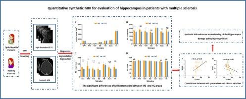
45 MS patients and 26 healthy controls (HCs) underwent synthetic MRI and 3D-T1 MRI. The hippocampus volumes were assessed by using voxel-based morphometry. Synthetic MRI parameters (T1, T2, and proton density (PD)) from hippocampus and its subfield were measured and compared, and their associations with the Expanded Disability Status Scale (EDSS), Symbol Digit Modalities Test (SDMT) scores were further investigated.

Results

There was no significant difference in hippocampal volume between MS patients and HCs. Compared with HCs, the T1, T2 and PD values of hippocampus and its subfield increased in MS patients. T2 values showed positive correlation with EDSS and negative correlation with SDMT.

Conclusions

Synthetic MRI can detect subtle quantitative changes of the hippocampus in MS patients with normal hippocampal volume. Specifically, Synthetic MRI parameters may apply as potentially effective imaging biomarker for hippocampus evaluation.
Abstract Image

用于评估多发性硬化症患者海马体的定量合成磁共振成像。
目的方法:45 名多发性硬化症患者和 26 名健康对照者(HCs)接受了合成 MRI 和 3D-T1 MRI 检查。方法:45 名多发性硬化症患者和 26 名健康对照者(HC)接受了合成 MRI 和三维-T1 MRI 检查,并使用基于体素的形态测量法评估了海马体积。对海马及其子野的合成 MRI 参数(T1、T2 和质子密度 (PD))进行了测量和比较,并进一步研究了它们与残疾状况扩展量表 (EDSS)、符号数字模型测试 (SDMT) 评分之间的关系:结果:多发性硬化症患者的海马体积与普通人无明显差异。与普通人相比,多发性硬化症患者海马及其亚区的 T1、T2 和 PD 值均有所增加。T2值与EDSS呈正相关,与SDMT呈负相关:结论:合成 MRI 可以检测海马体积正常的多发性硬化症患者海马的细微定量变化。具体而言,合成 MRI 参数可作为评估海马的潜在有效成像生物标志物。
本文章由计算机程序翻译,如有差异,请以英文原文为准。
求助全文
约1分钟内获得全文 求助全文
来源期刊
Brain Research
Brain Research 医学-神经科学
CiteScore
5.90
自引率
3.40%
发文量
268
审稿时长
47 days
期刊介绍: An international multidisciplinary journal devoted to fundamental research in the brain sciences. Brain Research publishes papers reporting interdisciplinary investigations of nervous system structure and function that are of general interest to the international community of neuroscientists. As is evident from the journals name, its scope is broad, ranging from cellular and molecular studies through systems neuroscience, cognition and disease. Invited reviews are also published; suggestions for and inquiries about potential reviews are welcomed. With the appearance of the final issue of the 2011 subscription, Vol. 67/1-2 (24 June 2011), Brain Research Reviews has ceased publication as a distinct journal separate from Brain Research. Review articles accepted for Brain Research are now published in that journal.
×
引用
GB/T 7714-2015
复制
MLA
复制
APA
复制
导出至
BibTeX EndNote RefMan NoteFirst NoteExpress
×
提示
您的信息不完整,为了账户安全,请先补充。
现在去补充
×
提示
您因"违规操作"
具体请查看互助需知
我知道了
×
提示
确定
请完成安全验证×
copy
已复制链接
快去分享给好友吧!
我知道了
右上角分享
点击右上角分享
0
联系我们:info@booksci.cn Book学术提供免费学术资源搜索服务,方便国内外学者检索中英文文献。致力于提供最便捷和优质的服务体验。 Copyright © 2023 布克学术 All rights reserved.
京ICP备2023020795号-1
ghs 京公网安备 11010802042870号
Book学术文献互助
Book学术文献互助群
群 号:481959085
Book学术官方微信