Pharmacological Inhibition of the Spliceosome SF3b Complex by Pladienolide-B Elicits Craniofacial Developmental Defects in Mouse and Zebrafish

IF 1.6 4区 医学 Q4 DEVELOPMENTAL BIOLOGY
Yukiko Hoshino, Shujie Liu, Toshiko Furutera, Takahiko Yamada, Daisuke Koyabu, Yuko Nukada, Masaaki Miyazawa, Tetsuya Yoda, Koichiro Ichimura, Sachiko Iseki, Junichi Tasaki, Masaki Takechi
{"title":"Pharmacological Inhibition of the Spliceosome SF3b Complex by Pladienolide-B Elicits Craniofacial Developmental Defects in Mouse and Zebrafish","authors":"Yukiko Hoshino,&nbsp;Shujie Liu,&nbsp;Toshiko Furutera,&nbsp;Takahiko Yamada,&nbsp;Daisuke Koyabu,&nbsp;Yuko Nukada,&nbsp;Masaaki Miyazawa,&nbsp;Tetsuya Yoda,&nbsp;Koichiro Ichimura,&nbsp;Sachiko Iseki,&nbsp;Junichi Tasaki,&nbsp;Masaki Takechi","doi":"10.1002/bdr2.2404","DOIUrl":null,"url":null,"abstract":"<div>\n \n \n <section>\n \n <h3> Background</h3>\n \n <p>Mutations in genes encoding spliceosome components result in craniofacial structural defects in humans, referred to as spliceosomopathies. The SF3b complex is a crucial unit of the spliceosome, but model organisms generated through genetic modification of the complex do not perfectly mimic the phenotype of spliceosomopathies. Since the phenotypes are suggested to be determined by the extent of spliceosome dysfunction, an alternative experimental system that can seamlessly control SF3b function is needed.</p>\n </section>\n \n <section>\n \n <h3> Methods</h3>\n \n <p>To establish another experimental system for model organisms elucidating relationship between spliceosome function and human diseases, we administered Pladienolide-B (PB), a SF3b complex inhibitor, to mouse and zebrafish embryos and assessed resulting phenotypes.</p>\n </section>\n \n <section>\n \n <h3> Results</h3>\n \n <p>PB-treated mouse embryos exhibited neural tube defect and exencephaly, accompanied by apoptosis and reduced cell proliferation in the neural tube, but normal structure in the midface and jaw. PB administration to heterozygous knockout mice of <i>Sf3b4</i>, a gene coding for a SF3b component, influenced the formation of cranial neural crest cells (CNCCs). Despite challenges in continuous PB administration and a high death rate in mice, PB was stably administered to zebrafish embryos, resulting in prolonged survival. Brain, cranial nerve, retina, midface, and jaw development were affected, mimicking spliceosomopathy phenotypes. Additionally, alterations in cell proliferation, cell death, and migration of CNCCs were detected.</p>\n </section>\n \n <section>\n \n <h3> Conclusions</h3>\n \n <p>We demonstrated that zebrafish treated with PB exhibited phenotypes similar to those observed in human spliceosomopathies. This experimental system may serve as a valuable research tool for understanding spliceosome function and human diseases.</p>\n </section>\n </div>","PeriodicalId":9121,"journal":{"name":"Birth Defects Research","volume":null,"pages":null},"PeriodicalIF":1.6000,"publicationDate":"2024-11-04","publicationTypes":"Journal Article","fieldsOfStudy":null,"isOpenAccess":false,"openAccessPdf":"https://onlinelibrary.wiley.com/doi/epdf/10.1002/bdr2.2404","citationCount":"0","resultStr":null,"platform":"Semanticscholar","paperid":null,"PeriodicalName":"Birth Defects Research","FirstCategoryId":"3","ListUrlMain":"https://onlinelibrary.wiley.com/doi/10.1002/bdr2.2404","RegionNum":4,"RegionCategory":"医学","ArticlePicture":[],"TitleCN":null,"AbstractTextCN":null,"PMCID":null,"EPubDate":"","PubModel":"","JCR":"Q4","JCRName":"DEVELOPMENTAL BIOLOGY","Score":null,"Total":0}
引用次数: 0

Abstract

Background

Mutations in genes encoding spliceosome components result in craniofacial structural defects in humans, referred to as spliceosomopathies. The SF3b complex is a crucial unit of the spliceosome, but model organisms generated through genetic modification of the complex do not perfectly mimic the phenotype of spliceosomopathies. Since the phenotypes are suggested to be determined by the extent of spliceosome dysfunction, an alternative experimental system that can seamlessly control SF3b function is needed.

Methods

To establish another experimental system for model organisms elucidating relationship between spliceosome function and human diseases, we administered Pladienolide-B (PB), a SF3b complex inhibitor, to mouse and zebrafish embryos and assessed resulting phenotypes.

Results

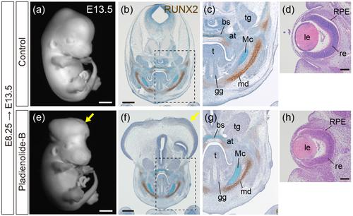
PB-treated mouse embryos exhibited neural tube defect and exencephaly, accompanied by apoptosis and reduced cell proliferation in the neural tube, but normal structure in the midface and jaw. PB administration to heterozygous knockout mice of Sf3b4, a gene coding for a SF3b component, influenced the formation of cranial neural crest cells (CNCCs). Despite challenges in continuous PB administration and a high death rate in mice, PB was stably administered to zebrafish embryos, resulting in prolonged survival. Brain, cranial nerve, retina, midface, and jaw development were affected, mimicking spliceosomopathy phenotypes. Additionally, alterations in cell proliferation, cell death, and migration of CNCCs were detected.

Conclusions

We demonstrated that zebrafish treated with PB exhibited phenotypes similar to those observed in human spliceosomopathies. This experimental system may serve as a valuable research tool for understanding spliceosome function and human diseases.
Abstract Image

Pladienolide-B 对剪接体 SF3b 复合物的药理抑制导致小鼠和斑马鱼颅面发育缺陷
背景:编码剪接体成分的基因突变导致人类颅面结构缺陷,被称为剪接体病。SF3b 复合物是剪接体的关键单元,但通过对该复合物进行基因修饰而产生的模式生物并不能完全模拟剪接体病的表型。由于表型是由剪接体功能障碍的程度决定的,因此需要一种能无缝控制 SF3b 功能的替代实验系统:方法:为了建立另一个实验系统,用于阐明剪接体功能与人类疾病之间关系的模式生物,我们给小鼠和斑马鱼胚胎注射了一种SF3b复合体抑制剂Pladienolide-B(PB),并评估了由此产生的表型:结果:经PB处理的小鼠胚胎表现出神经管缺陷和无脑畸形,并伴有神经管细胞凋亡和细胞增殖减少,但中面部和下颌结构正常。给Sf3b4(一种编码SF3b成分的基因)杂合子基因敲除小鼠注射PB会影响颅神经嵴细胞(CNCC)的形成。尽管持续给药 PB 存在挑战,而且小鼠的死亡率很高,但给斑马鱼胚胎稳定给药 PB 却能延长其存活时间。斑马鱼的大脑、颅神经、视网膜、中面部和下颌的发育受到影响,模仿了剪接体病的表型。此外,还检测到 CNCCs 的细胞增殖、细胞死亡和迁移发生了改变:我们证明了斑马鱼经 PB 处理后表现出与人类剪接体病相似的表型。这一实验系统可作为了解剪接体功能和人类疾病的重要研究工具。
本文章由计算机程序翻译,如有差异,请以英文原文为准。
求助全文
约1分钟内获得全文 求助全文
来源期刊
Birth Defects Research
Birth Defects Research Medicine-Embryology
CiteScore
3.60
自引率
9.50%
发文量
153
期刊介绍: The journal Birth Defects Research publishes original research and reviews in areas related to the etiology of adverse developmental and reproductive outcome. In particular the journal is devoted to the publication of original scientific research that contributes to the understanding of the biology of embryonic development and the prenatal causative factors and mechanisms leading to adverse pregnancy outcomes, namely structural and functional birth defects, pregnancy loss, postnatal functional defects in the human population, and to the identification of prenatal factors and biological mechanisms that reduce these risks. Adverse reproductive and developmental outcomes may have genetic, environmental, nutritional or epigenetic causes. Accordingly, the journal Birth Defects Research takes an integrated, multidisciplinary approach in its organization and publication strategy. The journal Birth Defects Research contains separate sections for clinical and molecular teratology, developmental and reproductive toxicology, and reviews in developmental biology to acknowledge and accommodate the integrative nature of research in this field. Each section has a dedicated editor who is a leader in his/her field and who has full editorial authority in his/her area.
×
引用
GB/T 7714-2015
复制
MLA
复制
APA
复制
导出至
BibTeX EndNote RefMan NoteFirst NoteExpress
×
提示
您的信息不完整,为了账户安全,请先补充。
现在去补充
×
提示
您因"违规操作"
具体请查看互助需知
我知道了
×
提示
确定
请完成安全验证×
copy
已复制链接
快去分享给好友吧!
我知道了
右上角分享
点击右上角分享
0
联系我们:info@booksci.cn Book学术提供免费学术资源搜索服务,方便国内外学者检索中英文文献。致力于提供最便捷和优质的服务体验。 Copyright © 2023 布克学术 All rights reserved.
京ICP备2023020795号-1
ghs 京公网安备 11010802042870号
Book学术文献互助
Book学术文献互助群
群 号:481959085
Book学术官方微信