Cervical cancer diagnosis model using spontaneous Raman and Coherent anti-Stokes Raman spectroscopy with artificial intelligence
IF 4.3
2区 化学
Q1 SPECTROSCOPY
Spectrochimica Acta Part A: Molecular and Biomolecular Spectroscopy
Pub Date : 2024-10-28
DOI:10.1016/j.saa.2024.125353
引用次数: 0
Abstract
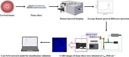
Cervical cancer is the fourth most common cancer worldwide. Histopathology, which is currently considered the gold standard for cervical cancer diagnosis, can be time-consuming and subjective. Therefore, there is an urgent need for a rapid, objective, and non-destructive cervical cancer detection technique. In this study, high-wavenumber spontaneous Raman spectroscopy was used to detect cervical squamous cell carcinoma and normal tissues. The levels of lipids, fatty acids, and proteins in cervical cancerous tissues were found to be higher than those in normal tissues. Raman difference spectroscopy revealed the most significant difference at 2928 cm−1. Additionally, a Coherent anti-Stokes Raman spectroscopy (CARS) instrument was employed to enhance the wavenumber signal intensity and sensitivity. The intrinsic relationship between CARS imaging and cervical lesions was established. The CARS images indicated that the intensity of normal cervical squamous cells was zero, whereas the intensities of keratinized and non-keratinized cervical squamous cell carcinoma tissues were significantly higher. Consequently, diagnostic outcomes could be obtained by observing CARS images with the naked eye. Furthermore, the characteristic structure of keratin pearls in keratinized cervical cancer could serve as a marker for subdividing cervical cancer types. Finally, a ConvNeXt network, a machine-learning model built from CARS images, was utilized to classify different types of tissue images. The results indicated a verification accuracy of 100 %, with a loss function of 0.0927. These findings suggest that the diagnostic model established using CARS images could efficiently diagnose cervical cancer, providing novel insights into the pathological diagnosis of this disease.

利用自发拉曼光谱和相干反斯托克斯拉曼光谱与人工智能的宫颈癌诊断模型
宫颈癌是全球第四大常见癌症。组织病理学目前被认为是诊断宫颈癌的黄金标准,但它既耗时又主观。因此,迫切需要一种快速、客观、非破坏性的宫颈癌检测技术。本研究采用高波长自发拉曼光谱检测宫颈鳞状细胞癌和正常组织。研究发现,宫颈癌组织中的脂质、脂肪酸和蛋白质含量高于正常组织。拉曼差异光谱显示,在 2928 cm-1 处差异最大。此外,还采用了相干反斯托克斯拉曼光谱(CARS)仪器来增强波长信号强度和灵敏度。建立了 CARS 成像与宫颈病变之间的内在联系。CARS 图像显示,正常宫颈鳞状细胞的强度为零,而角化和非角化宫颈鳞状细胞癌组织的强度则明显较高。因此,通过肉眼观察 CARS 图像可以获得诊断结果。此外,角质化宫颈癌中角蛋白珍珠的特征结构可作为细分宫颈癌类型的标记。最后,利用从 CARS 图像建立的机器学习模型 ConvNeXt 网络对不同类型的组织图像进行了分类。结果表明,验证准确率为 100%,损失函数为 0.0927。这些研究结果表明,利用 CARS 图像建立的诊断模型可以有效诊断宫颈癌,为该疾病的病理诊断提供了新的见解。
本文章由计算机程序翻译,如有差异,请以英文原文为准。
求助全文
约1分钟内获得全文
求助全文
来源期刊
CiteScore
8.40
自引率
11.40%
发文量
1364
审稿时长
40 days
期刊介绍:
Spectrochimica Acta, Part A: Molecular and Biomolecular Spectroscopy (SAA) is an interdisciplinary journal which spans from basic to applied aspects of optical spectroscopy in chemistry, medicine, biology, and materials science.
The journal publishes original scientific papers that feature high-quality spectroscopic data and analysis. From the broad range of optical spectroscopies, the emphasis is on electronic, vibrational or rotational spectra of molecules, rather than on spectroscopy based on magnetic moments.
Criteria for publication in SAA are novelty, uniqueness, and outstanding quality. Routine applications of spectroscopic techniques and computational methods are not appropriate.
Topics of particular interest of Spectrochimica Acta Part A include, but are not limited to:
Spectroscopy and dynamics of bioanalytical, biomedical, environmental, and atmospheric sciences,
Novel experimental techniques or instrumentation for molecular spectroscopy,
Novel theoretical and computational methods,
Novel applications in photochemistry and photobiology,
Novel interpretational approaches as well as advances in data analysis based on electronic or vibrational spectroscopy.

 求助内容:
求助内容: 应助结果提醒方式:


