Veronika Kulik, Melissa K. Edler, Mary Ann Raghanti, Aminu Imam, Chet C. Sherwood
{"title":"Amyloid-Beta, Tau, and Microglial Activation in Aged Felid Brains","authors":"Veronika Kulik, Melissa K. Edler, Mary Ann Raghanti, Aminu Imam, Chet C. Sherwood","doi":"10.1002/cne.25679","DOIUrl":null,"url":null,"abstract":"<div>\n \n <p>Alzheimer's disease (AD) and its associated pathology have been primarily identified in humans, who have relatively large brains and long lifespans. To expand what is known about aging and neurodegeneration across mammalian species, we characterized amyloid-beta (Aβ) and tau lesions in five species of aged felids (<i>n</i> = 9; cheetah, clouded leopard, African lion, serval, Siberian tiger). We performed immunohistochemistry to detect Aβ40 and Aβ42 in plaques and vessels and hyperphosphorylated tau in the temporal lobe gyrus sylvius and in the CA1 and CA3 subfields of the hippocampus. We also quantified the densities and morphological types of microglia expressing IBA1. We found that diffuse Aβ42 plaques, but not dense-core plaques, were present more frequently in the temporal cortex and tended to be more common than Aβ40 plaques across species. Conversely, vascular Aβ was labeled more consistently with Aβ40 for each species on average. Although all individuals showed some degree of Aβ40 and/or Aβ42 immunoreactivity, only the cheetahs and clouded leopards exhibited intraneuronal hyperphosphorylated tau (i.e., pretangles), which was more common in the hippocampus. Reactive, intermediate microglia were significantly associated with total Aβ40 vessel area and pretangle load in the hippocampus. This study demonstrates the co-occurrence of Aβ and tau pathology in two felid species, cheetahs and clouded leopards. Overall, these results provide an initial view of the manifestation of Aβ and tau pathology in aged, large-brained felids, which can be compared with markers of neurodegeneration across different taxa, including domestic cats, nonhuman primates, and humans.</p>\n </div>","PeriodicalId":15552,"journal":{"name":"Journal of Comparative Neurology","volume":"532 11","pages":""},"PeriodicalIF":2.1000,"publicationDate":"2024-10-30","publicationTypes":"Journal Article","fieldsOfStudy":null,"isOpenAccess":false,"openAccessPdf":"","citationCount":"0","resultStr":null,"platform":"Semanticscholar","paperid":null,"PeriodicalName":"Journal of Comparative Neurology","FirstCategoryId":"3","ListUrlMain":"https://onlinelibrary.wiley.com/doi/10.1002/cne.25679","RegionNum":4,"RegionCategory":"医学","ArticlePicture":[],"TitleCN":null,"AbstractTextCN":null,"PMCID":null,"EPubDate":"","PubModel":"","JCR":"Q3","JCRName":"NEUROSCIENCES","Score":null,"Total":0}
引用次数: 0
Abstract
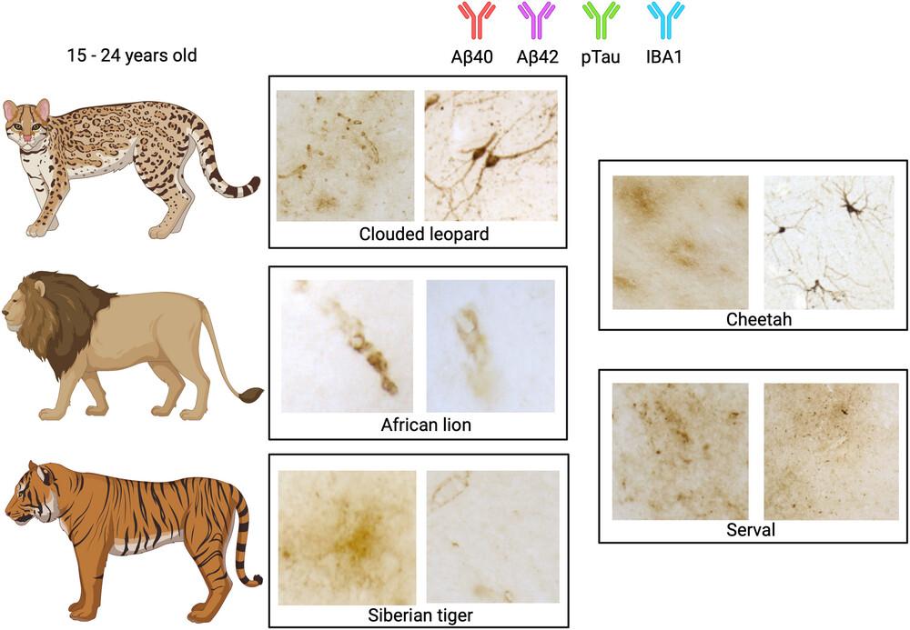
Alzheimer's disease (AD) and its associated pathology have been primarily identified in humans, who have relatively large brains and long lifespans. To expand what is known about aging and neurodegeneration across mammalian species, we characterized amyloid-beta (Aβ) and tau lesions in five species of aged felids (n = 9; cheetah, clouded leopard, African lion, serval, Siberian tiger). We performed immunohistochemistry to detect Aβ40 and Aβ42 in plaques and vessels and hyperphosphorylated tau in the temporal lobe gyrus sylvius and in the CA1 and CA3 subfields of the hippocampus. We also quantified the densities and morphological types of microglia expressing IBA1. We found that diffuse Aβ42 plaques, but not dense-core plaques, were present more frequently in the temporal cortex and tended to be more common than Aβ40 plaques across species. Conversely, vascular Aβ was labeled more consistently with Aβ40 for each species on average. Although all individuals showed some degree of Aβ40 and/or Aβ42 immunoreactivity, only the cheetahs and clouded leopards exhibited intraneuronal hyperphosphorylated tau (i.e., pretangles), which was more common in the hippocampus. Reactive, intermediate microglia were significantly associated with total Aβ40 vessel area and pretangle load in the hippocampus. This study demonstrates the co-occurrence of Aβ and tau pathology in two felid species, cheetahs and clouded leopards. Overall, these results provide an initial view of the manifestation of Aβ and tau pathology in aged, large-brained felids, which can be compared with markers of neurodegeneration across different taxa, including domestic cats, nonhuman primates, and humans.
期刊介绍:
Established in 1891, JCN is the oldest continually published basic neuroscience journal. Historically, as the name suggests, the journal focused on a comparison among species to uncover the intricacies of how the brain functions. In modern times, this research is called systems neuroscience where animal models are used to mimic core cognitive processes with the ultimate goal of understanding neural circuits and connections that give rise to behavioral patterns and different neural states.
Research published in JCN covers all species from invertebrates to humans, and the reports inform the readers about the function and organization of nervous systems in species with an emphasis on the way that species adaptations inform about the function or organization of the nervous systems, rather than on their evolution per se.
JCN publishes primary research articles and critical commentaries and review-type articles offering expert insight in to cutting edge research in the field of systems neuroscience; a complete list of contribution types is given in the Author Guidelines. For primary research contributions, only full-length investigative reports are desired; the journal does not accept short communications.

 求助内容:
求助内容: 应助结果提醒方式:


