Assessment of submicron bone tissue composition in plastic-embedded samples using optical photothermal infrared (O-PTIR) spectral imaging and machine learning

IF 3.5 Q2 BIOCHEMISTRY & MOLECULAR BIOLOGY
Isha Dev , Sofia Mehmood , Nancy Pleshko , Iyad Obeid , William Querido
{"title":"Assessment of submicron bone tissue composition in plastic-embedded samples using optical photothermal infrared (O-PTIR) spectral imaging and machine learning","authors":"Isha Dev ,&nbsp;Sofia Mehmood ,&nbsp;Nancy Pleshko ,&nbsp;Iyad Obeid ,&nbsp;William Querido","doi":"10.1016/j.yjsbx.2024.100111","DOIUrl":null,"url":null,"abstract":"<div><div>Understanding the composition of bone tissue at the submicron level is crucial to elucidate factors contributing to bone disease and fragility. Here, we introduce a novel approach utilizing optical photothermal infrared (O-PTIR) spectroscopy and imaging coupled with machine learning analysis to assess bone tissue composition at 500 nm spatial resolution. This approach was used to evaluate thick bone samples embedded in typical poly(methyl methacrylate) (PMMA) blocks, eliminating the need for cumbersome thin sectioning. We demonstrate the utility of O-PTIR imaging to assess the distribution of bone tissue mineral and protein, as well as to explore the structure-composition relationship surrounding microporosity at a spatial resolution unattainable by conventional infrared imaging modalities. Using bone samples from wildtype (WT) mice and from a mouse model of osteogenesis imperfecta (OIM), we further showcase the application of O-PTIR spectroscopy to quantify mineral content, crystallinity, and carbonate content in spatially defined regions across the cortical bone. Notably, we show that machine learning analysis using support vector machine (SVM) was successful in identifying bone phenotypes (typical in WT, fragile in OIM) based on input of spectral data, with over 86 % of samples correctly identified when using the collagen spectral range. Our findings highlight the potential of O-PTIR spectroscopy and imaging as valuable tools for exploring bone submicron composition.</div></div>","PeriodicalId":17238,"journal":{"name":"Journal of Structural Biology: X","volume":"10 ","pages":"Article 100111"},"PeriodicalIF":3.5000,"publicationDate":"2024-10-09","publicationTypes":"Journal Article","fieldsOfStudy":null,"isOpenAccess":false,"openAccessPdf":"","citationCount":"0","resultStr":null,"platform":"Semanticscholar","paperid":null,"PeriodicalName":"Journal of Structural Biology: X","FirstCategoryId":"1085","ListUrlMain":"https://www.sciencedirect.com/science/article/pii/S2590152424000163","RegionNum":0,"RegionCategory":null,"ArticlePicture":[],"TitleCN":null,"AbstractTextCN":null,"PMCID":null,"EPubDate":"","PubModel":"","JCR":"Q2","JCRName":"BIOCHEMISTRY & MOLECULAR BIOLOGY","Score":null,"Total":0}
引用次数: 0

Abstract

Understanding the composition of bone tissue at the submicron level is crucial to elucidate factors contributing to bone disease and fragility. Here, we introduce a novel approach utilizing optical photothermal infrared (O-PTIR) spectroscopy and imaging coupled with machine learning analysis to assess bone tissue composition at 500 nm spatial resolution. This approach was used to evaluate thick bone samples embedded in typical poly(methyl methacrylate) (PMMA) blocks, eliminating the need for cumbersome thin sectioning. We demonstrate the utility of O-PTIR imaging to assess the distribution of bone tissue mineral and protein, as well as to explore the structure-composition relationship surrounding microporosity at a spatial resolution unattainable by conventional infrared imaging modalities. Using bone samples from wildtype (WT) mice and from a mouse model of osteogenesis imperfecta (OIM), we further showcase the application of O-PTIR spectroscopy to quantify mineral content, crystallinity, and carbonate content in spatially defined regions across the cortical bone. Notably, we show that machine learning analysis using support vector machine (SVM) was successful in identifying bone phenotypes (typical in WT, fragile in OIM) based on input of spectral data, with over 86 % of samples correctly identified when using the collagen spectral range. Our findings highlight the potential of O-PTIR spectroscopy and imaging as valuable tools for exploring bone submicron composition.
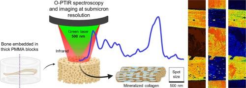
Abstract Image

利用光学光热红外(O-PTIR)光谱成像和机器学习评估塑料包埋样本中的亚微米骨组织成分
了解亚微米级的骨组织成分对于阐明导致骨病和骨脆性的因素至关重要。在这里,我们介绍了一种利用光学光热红外(O-PTIR)光谱和成像并结合机器学习分析的新方法,以 500 纳米的空间分辨率评估骨组织成分。这种方法可用于评估嵌入典型聚甲基丙烯酸甲酯(PMMA)块中的厚骨样本,从而省去了繁琐的薄切片检查。我们展示了 O-PTIR 成像在评估骨组织矿物质和蛋白质分布方面的实用性,以及在传统红外成像模式无法达到的空间分辨率下探索围绕微孔的结构-组成关系。利用野生型(WT)小鼠和成骨不全症(OIM)小鼠模型的骨样本,我们进一步展示了如何应用 O-PTIR 光谱量化整个皮质骨空间定义区域的矿物质含量、结晶度和碳酸盐含量。值得注意的是,我们展示了使用支持向量机(SVM)进行的机器学习分析能够根据输入的光谱数据成功识别骨表型(WT 中典型,OIM 中脆弱),在使用胶原蛋白光谱范围时,超过 86% 的样本被正确识别。我们的研究结果凸显了 O-PTIR 光谱和成像作为探索骨亚微米组成的宝贵工具的潜力。
本文章由计算机程序翻译,如有差异,请以英文原文为准。
求助全文
约1分钟内获得全文 求助全文
来源期刊
Journal of Structural Biology: X
Journal of Structural Biology: X Biochemistry, Genetics and Molecular Biology-Structural Biology
CiteScore
6.50
自引率
0.00%
发文量
20
审稿时长
62 days
×
引用
GB/T 7714-2015
复制
MLA
复制
APA
复制
导出至
BibTeX EndNote RefMan NoteFirst NoteExpress
×
提示
您的信息不完整,为了账户安全,请先补充。
现在去补充
×
提示
您因"违规操作"
具体请查看互助需知
我知道了
×
提示
确定
请完成安全验证×
copy
已复制链接
快去分享给好友吧!
我知道了
右上角分享
点击右上角分享
0
联系我们:info@booksci.cn Book学术提供免费学术资源搜索服务,方便国内外学者检索中英文文献。致力于提供最便捷和优质的服务体验。 Copyright © 2023 布克学术 All rights reserved.
京ICP备2023020795号-1
ghs 京公网安备 11010802042870号
Book学术文献互助
Book学术文献互助群
群 号:481959085
Book学术官方微信