Mari's novel T shaped incision in frenotomy technique with bilateral pedicle flap- an aesthetic approach
Q1 Medicine
Journal of oral biology and craniofacial research
Pub Date : 2024-10-22
DOI:10.1016/j.jobcr.2024.09.008
引用次数: 0
Abstract
Background
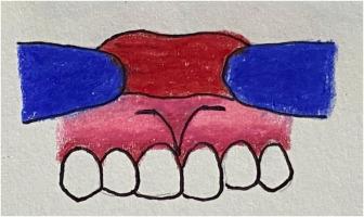
Aberrant frenum attachments often lead to mucogingival deformities, culminating in both functional impairments and aesthetic concerns. Traditional frenectomy procedures are associated with extensive incisions and resultant wound defects. To address these challenges, a novel T-shaped incision technique has been developed. This method enhances accessibility for removing vestibular and frenal attachments while creating a tripod layer of flaps, thereby promoting primary closure and improved blood supply for healing by primary intention.
Case management
This innovative technique is particularly suited for managing frenum variations such as bifid frenum, persistent tectolabial frenum, double frenum, and wider frenum. The procedure involves crafting two horizontal and two converging parallel incisions that form a “T" shape, facilitating effective fiber detachment. Bilateral pedicle flaps are then employed for primary closure, which concurrently enlarges the zone of attached gingiva.
Conclusion
The T-shaped incision approach presents an advanced method for treating abnormal frenum attachments, ensuring efficient fiber separation and fostering healing by primary intention.

Mari 在双侧椎弓根皮瓣腱索切开术中采用新颖的 T 形切口--一种美学方法
背景尖锐的龈缘附着常常导致粘牙龈畸形,最终造成功能障碍和美观问题。传统的龈沟切除术切口大,会造成伤口缺损。为了应对这些挑战,我们开发了一种新颖的 T 形切口技术。这种方法提高了切除前庭和蹼附着物的可及性,同时创建了一个三脚架皮瓣层,从而促进原发闭合,改善血液供应,促进原发意向愈合。这种创新技术尤其适用于处理蹼的变异,如二叉蹼、持续性齿状蹼、双蹼和较宽的蹼。手术过程包括制作两个水平切口和两个会聚的平行切口,形成 "T "形,以便有效分离纤维。结论 "T "形切口法是治疗异常龈缘附着的一种先进方法,可确保有效的纤维分离并促进原发意向愈合。
本文章由计算机程序翻译,如有差异,请以英文原文为准。
求助全文
约1分钟内获得全文
求助全文
来源期刊

Journal of oral biology and craniofacial research
Medicine-Otorhinolaryngology
CiteScore
4.90
自引率
0.00%
发文量
133
审稿时长
167 days
期刊介绍:
Journal of Oral Biology and Craniofacial Research (JOBCR)is the official journal of the Craniofacial Research Foundation (CRF). The journal aims to provide a common platform for both clinical and translational research and to promote interdisciplinary sciences in craniofacial region. JOBCR publishes content that includes diseases, injuries and defects in the head, neck, face, jaws and the hard and soft tissues of the mouth and jaws and face region; diagnosis and medical management of diseases specific to the orofacial tissues and of oral manifestations of systemic diseases; studies on identifying populations at risk of oral disease or in need of specific care, and comparing regional, environmental, social, and access similarities and differences in dental care between populations; diseases of the mouth and related structures like salivary glands, temporomandibular joints, facial muscles and perioral skin; biomedical engineering, tissue engineering and stem cells. The journal publishes reviews, commentaries, peer-reviewed original research articles, short communication, and case reports.
 求助内容:
求助内容: 应助结果提醒方式:


