Raúl Gijón Villanova, Concepción López Peña, Alfonso Extremera Ortega, Alicia Martín-Lagos Maldonado, José Miguel Candel Erenas
{"title":"Gastric cancer of unusual presentation, the importance of differential diagnosis.","authors":"Raúl Gijón Villanova, Concepción López Peña, Alfonso Extremera Ortega, Alicia Martín-Lagos Maldonado, José Miguel Candel Erenas","doi":"10.17235/reed.2024.10725/2024","DOIUrl":null,"url":null,"abstract":"<p><p>We present the case of a 67-year-old male smoker with no medical history of interest. Admitted to Neurology for frontal headache, unsteady gait, temporospatial disorientation and vomiting. Laboratory tests (including vitamin B12, folic acid, lues) and cranial CT scan were normal, encephalogram compatible with diffuse encephalopathy and lumbar puncture with a finding of leptomeningeal carcinomatosis. In light of these findings, it was decided to look for occult tumor by thoracoabdominal-pelvic CT, which was negative for malignancy. In view of the results of the previous tests, it was decided to perform an endoscopic study. Colonoscopy reveals six 0-IIa Paris polyps in the left colon measuring 7-10 cm, which are removed. Gastroduodenoscopy shows a poorly distensible stomach, with erythematous gastric body mucosa and hard on biopsy. In addition, in the duodenum, three raised lesions with an excavated center (Fig. 1) of about 5 mm were identified and biopsied. Histology findings report mucosal and submucosal infiltration by poorly differentiated carcinoma. Immunohistochemistry positive for CK19, Glipican-3, weak positivity for CK7, conserved expression of MUC5AC and SMAD, negative for SF-1, inhibin, synaptophysin, INSM1, chromogramin, Gata-3 and S-100. The findings were suggestive of infiltration by poorly differentiated carcinoma of probable gastric origin. Unfortunately, during hospital admission the patient presented a progressive clinical deterioration and died two weeks later.</p>","PeriodicalId":21342,"journal":{"name":"Revista Espanola De Enfermedades Digestivas","volume":" ","pages":""},"PeriodicalIF":2.7000,"publicationDate":"2024-10-18","publicationTypes":"Journal Article","fieldsOfStudy":null,"isOpenAccess":false,"openAccessPdf":"","citationCount":"0","resultStr":null,"platform":"Semanticscholar","paperid":null,"PeriodicalName":"Revista Espanola De Enfermedades Digestivas","FirstCategoryId":"3","ListUrlMain":"https://doi.org/10.17235/reed.2024.10725/2024","RegionNum":4,"RegionCategory":"医学","ArticlePicture":[],"TitleCN":null,"AbstractTextCN":null,"PMCID":null,"EPubDate":"","PubModel":"","JCR":"Q2","JCRName":"GASTROENTEROLOGY & HEPATOLOGY","Score":null,"Total":0}
引用次数: 0
Abstract
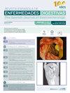
We present the case of a 67-year-old male smoker with no medical history of interest. Admitted to Neurology for frontal headache, unsteady gait, temporospatial disorientation and vomiting. Laboratory tests (including vitamin B12, folic acid, lues) and cranial CT scan were normal, encephalogram compatible with diffuse encephalopathy and lumbar puncture with a finding of leptomeningeal carcinomatosis. In light of these findings, it was decided to look for occult tumor by thoracoabdominal-pelvic CT, which was negative for malignancy. In view of the results of the previous tests, it was decided to perform an endoscopic study. Colonoscopy reveals six 0-IIa Paris polyps in the left colon measuring 7-10 cm, which are removed. Gastroduodenoscopy shows a poorly distensible stomach, with erythematous gastric body mucosa and hard on biopsy. In addition, in the duodenum, three raised lesions with an excavated center (Fig. 1) of about 5 mm were identified and biopsied. Histology findings report mucosal and submucosal infiltration by poorly differentiated carcinoma. Immunohistochemistry positive for CK19, Glipican-3, weak positivity for CK7, conserved expression of MUC5AC and SMAD, negative for SF-1, inhibin, synaptophysin, INSM1, chromogramin, Gata-3 and S-100. The findings were suggestive of infiltration by poorly differentiated carcinoma of probable gastric origin. Unfortunately, during hospital admission the patient presented a progressive clinical deterioration and died two weeks later.
期刊介绍:
La Revista Española de Enfermedades Digestivas, Órgano Oficial de la Sociedad Española de Patología Digestiva (SEPD), Sociedad Española de Endoscopia Digestiva (SEED) y Asociación Española de Ecografía Digestiva (AEED), publica artículos originales, editoriales, revisiones, casos clínicos, cartas al director, imágenes en patología digestiva, y otros artículos especiales sobre todos los aspectos relativos a las enfermedades digestivas.
 求助内容:
求助内容: 应助结果提醒方式:


