Brea Chernokal, Bryan J. Ferrick and Jason P. Gleghorn
{"title":"Zonal patterning of extracellular matrix and stromal cell populations along a perfusable cellular microchannel†","authors":"Brea Chernokal, Bryan J. Ferrick and Jason P. Gleghorn","doi":"10.1039/D4LC00579A","DOIUrl":null,"url":null,"abstract":"<p >The spatial organization of biophysical and biochemical cues in the extracellular matrix (ECM) in concert with reciprocal cell–cell signaling is vital to tissue patterning during development. However, elucidating the role an individual microenvironmental factor plays using existing <em>in vivo</em> models is difficult due to their inherent complexity. In this work, we have developed a microphysiological system to spatially pattern the biochemical, biophysical, and stromal cell composition of the ECM along an epithelialized 3D microchannel. This technique is adaptable to multiple hydrogel compositions and scalable to the number of zones patterned. We confirmed that the methodology to create distinct zones resulted in a continuous, annealed hydrogel with regional interfaces that did not hinder the transport of soluble molecules. Further, the interface between hydrogel regions did not disrupt microchannel structure, epithelial lumen formation, or media perfusion through an acellular or cellularized microchannel. Finally, we demonstrated spatially patterned tubulogenic sprouting of a continuous epithelial tube into the surrounding hydrogel confined to local regions with stromal cell populations, illustrating spatial control of cell–cell interactions and signaling gradients. This easy-to-use system has wide utility for modeling three-dimensional epithelial and endothelial tissue interactions with heterogeneous hydrogel compositions and/or stromal cell populations to investigate their mechanistic roles during development, homeostasis, or disease.</p>","PeriodicalId":85,"journal":{"name":"Lab on a Chip","volume":" 23","pages":" 5238-5250"},"PeriodicalIF":6.1000,"publicationDate":"2024-10-21","publicationTypes":"Journal Article","fieldsOfStudy":null,"isOpenAccess":false,"openAccessPdf":"https://pubs.rsc.org/en/content/articlepdf/2024/lc/d4lc00579a?page=search","citationCount":"0","resultStr":null,"platform":"Semanticscholar","paperid":null,"PeriodicalName":"Lab on a Chip","FirstCategoryId":"5","ListUrlMain":"https://pubs.rsc.org/en/content/articlelanding/2024/lc/d4lc00579a","RegionNum":2,"RegionCategory":"工程技术","ArticlePicture":[],"TitleCN":null,"AbstractTextCN":null,"PMCID":null,"EPubDate":"","PubModel":"","JCR":"Q1","JCRName":"BIOCHEMICAL RESEARCH METHODS","Score":null,"Total":0}
引用次数: 0
Abstract
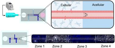
The spatial organization of biophysical and biochemical cues in the extracellular matrix (ECM) in concert with reciprocal cell–cell signaling is vital to tissue patterning during development. However, elucidating the role an individual microenvironmental factor plays using existing in vivo models is difficult due to their inherent complexity. In this work, we have developed a microphysiological system to spatially pattern the biochemical, biophysical, and stromal cell composition of the ECM along an epithelialized 3D microchannel. This technique is adaptable to multiple hydrogel compositions and scalable to the number of zones patterned. We confirmed that the methodology to create distinct zones resulted in a continuous, annealed hydrogel with regional interfaces that did not hinder the transport of soluble molecules. Further, the interface between hydrogel regions did not disrupt microchannel structure, epithelial lumen formation, or media perfusion through an acellular or cellularized microchannel. Finally, we demonstrated spatially patterned tubulogenic sprouting of a continuous epithelial tube into the surrounding hydrogel confined to local regions with stromal cell populations, illustrating spatial control of cell–cell interactions and signaling gradients. This easy-to-use system has wide utility for modeling three-dimensional epithelial and endothelial tissue interactions with heterogeneous hydrogel compositions and/or stromal cell populations to investigate their mechanistic roles during development, homeostasis, or disease.
期刊介绍:
Lab on a Chip is the premiere journal that publishes cutting-edge research in the field of miniaturization. By their very nature, microfluidic/nanofluidic/miniaturized systems are at the intersection of disciplines, spanning fundamental research to high-end application, which is reflected by the broad readership of the journal. Lab on a Chip publishes two types of papers on original research: full-length research papers and communications. Papers should demonstrate innovations, which can come from technical advancements or applications addressing pressing needs in globally important areas. The journal also publishes Comments, Reviews, and Perspectives.
 求助内容:
求助内容: 应助结果提醒方式:


