Forkhead box C1 promotes the pathology of osteoarthritis in subchondral bone osteoblasts via the Piezo1/YAP axis

IF 4.4 2区 生物学 Q2 CELL BIOLOGY
Zhengyuan Li , Lin Hao , Shenghong Chen , Wenhan Fu , Hui Zhang , Zongsheng Yin , Yin Wang , Jun Wang
{"title":"Forkhead box C1 promotes the pathology of osteoarthritis in subchondral bone osteoblasts via the Piezo1/YAP axis","authors":"Zhengyuan Li ,&nbsp;Lin Hao ,&nbsp;Shenghong Chen ,&nbsp;Wenhan Fu ,&nbsp;Hui Zhang ,&nbsp;Zongsheng Yin ,&nbsp;Yin Wang ,&nbsp;Jun Wang","doi":"10.1016/j.cellsig.2024.111463","DOIUrl":null,"url":null,"abstract":"<div><div>Subchondral bone sclerosis is a key characteristic of osteoarthritis (OA). Prior research has shown that Forkhead box C1 (FoxC1) plays a role in the synovial inflammation of OA, but its specific role in the subchondral bone of OA has not been explored. Our research revealed elevated expression levels of FoxC1 and Piezo1 in OA subchondral bone tissues. Further experiments on OA subchondral bone osteoblasts with FoxC1 or Piezo1 overexpression showed increased cell proliferation activity, expression of Yes-associated Protein 1 (YAP) and osteogenic markers, and secretion of proinflammatory factors. Mechanistically, the overexpression of FoxC1 through Piezo1 activation, in combination with downstream YAP signaling, led to increased levels of alkaline phosphatase (ALP), collagen type 1 (COL1) A1, RUNX2, Osteocalcin, matrix metalloproteinase (MMP) 3, and MMP9 expression. Notably, inhibition of Piezo1 reversed the regulatory function of FoxC1. The binding of FoxC1 to the targeted area (ATATTTATTTA, residues +612 to +622) and the activation of Piezo1 transcription were verified by the dual luciferase assays. Additionally, Reduced subchondral osteosclerosis and microangiogenesis were observed in knee joints from FoxC1-conditional knockout (CKO) and Piezo1-CKO mice, indicating reduced lesions. Collectively, our study reveals the significant involvement of FoxC1 in the pathologic process of OA subchondral bone via the Piezo1/YAP signaling pathway, potentially establishing a novel therapeutic target.</div></div>","PeriodicalId":9902,"journal":{"name":"Cellular signalling","volume":"124 ","pages":"Article 111463"},"PeriodicalIF":4.4000,"publicationDate":"2024-10-11","publicationTypes":"Journal Article","fieldsOfStudy":null,"isOpenAccess":false,"openAccessPdf":"","citationCount":"0","resultStr":null,"platform":"Semanticscholar","paperid":null,"PeriodicalName":"Cellular signalling","FirstCategoryId":"99","ListUrlMain":"https://www.sciencedirect.com/science/article/pii/S0898656824004364","RegionNum":2,"RegionCategory":"生物学","ArticlePicture":[],"TitleCN":null,"AbstractTextCN":null,"PMCID":null,"EPubDate":"","PubModel":"","JCR":"Q2","JCRName":"CELL BIOLOGY","Score":null,"Total":0}
引用次数: 0

Abstract

Subchondral bone sclerosis is a key characteristic of osteoarthritis (OA). Prior research has shown that Forkhead box C1 (FoxC1) plays a role in the synovial inflammation of OA, but its specific role in the subchondral bone of OA has not been explored. Our research revealed elevated expression levels of FoxC1 and Piezo1 in OA subchondral bone tissues. Further experiments on OA subchondral bone osteoblasts with FoxC1 or Piezo1 overexpression showed increased cell proliferation activity, expression of Yes-associated Protein 1 (YAP) and osteogenic markers, and secretion of proinflammatory factors. Mechanistically, the overexpression of FoxC1 through Piezo1 activation, in combination with downstream YAP signaling, led to increased levels of alkaline phosphatase (ALP), collagen type 1 (COL1) A1, RUNX2, Osteocalcin, matrix metalloproteinase (MMP) 3, and MMP9 expression. Notably, inhibition of Piezo1 reversed the regulatory function of FoxC1. The binding of FoxC1 to the targeted area (ATATTTATTTA, residues +612 to +622) and the activation of Piezo1 transcription were verified by the dual luciferase assays. Additionally, Reduced subchondral osteosclerosis and microangiogenesis were observed in knee joints from FoxC1-conditional knockout (CKO) and Piezo1-CKO mice, indicating reduced lesions. Collectively, our study reveals the significant involvement of FoxC1 in the pathologic process of OA subchondral bone via the Piezo1/YAP signaling pathway, potentially establishing a novel therapeutic target.
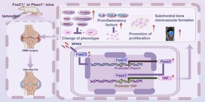
Abstract Image

叉头盒 C1 通过 Piezo1/YAP 轴促进软骨下骨成骨细胞骨关节炎的病理变化
软骨下骨硬化是骨关节炎(OA)的一个主要特征。先前的研究表明,叉头盒 C1(FoxC1)在 OA 的滑膜炎症中发挥作用,但其在 OA 软骨下骨中的具体作用尚未得到探讨。我们的研究发现,FoxC1 和 Piezo1 在 OA 软骨下骨组织中的表达水平升高。对 FoxC1 或 Piezo1 过表达的 OA 软骨下骨成骨细胞的进一步实验显示,细胞增殖活性、Yes 相关蛋白 1(YAP)和成骨标志物的表达以及促炎因子的分泌均有所增加。从机理上讲,通过激活 Piezo1 过表达 FoxC1 与下游 YAP 信号结合,导致碱性磷酸酶(ALP)、1 型胶原蛋白(COL1)A1、RUNX2、骨钙蛋白、基质金属蛋白酶(MMP)3 和 MMP9 表达水平升高。值得注意的是,抑制 Piezo1 会逆转 FoxC1 的调控功能。双荧光素酶试验验证了 FoxC1 与靶区(ATATTTATTTA,残基 +612 至 +622)的结合以及对 Piezo1 转录的激活。此外,在 FoxC1 条件性基因敲除(CKO)和 Piezo1-CKO 小鼠的膝关节中观察到软骨下骨质硬化和微血管生成减少,表明病变减轻。总之,我们的研究揭示了FoxC1通过Piezo1/YAP信号通路在OA软骨下骨病理过程中的重要参与作用,有可能建立一个新的治疗靶点。
本文章由计算机程序翻译,如有差异,请以英文原文为准。
求助全文
约1分钟内获得全文 求助全文
来源期刊
Cellular signalling
Cellular signalling 生物-细胞生物学
CiteScore
8.40
自引率
0.00%
发文量
250
审稿时长
27 days
期刊介绍: Cellular Signalling publishes original research describing fundamental and clinical findings on the mechanisms, actions and structural components of cellular signalling systems in vitro and in vivo. Cellular Signalling aims at full length research papers defining signalling systems ranging from microorganisms to cells, tissues and higher organisms.
×
引用
GB/T 7714-2015
复制
MLA
复制
APA
复制
导出至
BibTeX EndNote RefMan NoteFirst NoteExpress
×
提示
您的信息不完整,为了账户安全,请先补充。
现在去补充
×
提示
您因"违规操作"
具体请查看互助需知
我知道了
×
提示
确定
请完成安全验证×
copy
已复制链接
快去分享给好友吧!
我知道了
右上角分享
点击右上角分享
0
联系我们:info@booksci.cn Book学术提供免费学术资源搜索服务,方便国内外学者检索中英文文献。致力于提供最便捷和优质的服务体验。 Copyright © 2023 布克学术 All rights reserved.
京ICP备2023020795号-1
ghs 京公网安备 11010802042870号
Book学术文献互助
Book学术文献互助群
群 号:481959085
Book学术官方微信