Neurochemical Characterization of Dopaminoceptive Cells in Song Control Nuclei of Canaries and Their Activation During Song Production: A Multiplex Fluorescent In Situ Hybridization Study
Chelsea M. Haakenson, Jacques Balthazart, Jonathan W. VanRyzin, Ashley E. Marquardt, Sydney E. Ashton, Margaret M. McCarthy, Gregory F. Ball
{"title":"Neurochemical Characterization of Dopaminoceptive Cells in Song Control Nuclei of Canaries and Their Activation During Song Production: A Multiplex Fluorescent In Situ Hybridization Study","authors":"Chelsea M. Haakenson, Jacques Balthazart, Jonathan W. VanRyzin, Ashley E. Marquardt, Sydney E. Ashton, Margaret M. McCarthy, Gregory F. Ball","doi":"10.1002/cne.25675","DOIUrl":null,"url":null,"abstract":"<p>Highly sensitive in situ hybridization procedures (RNAScope) were used to quantify the expression of three dopamine receptors (Drd1, Drd2, and Drd3) in two song control nuclei (HVC and the Area X of the basal ganglia) that are known to receive dopaminergic inputs and in the periaqueductal gray (PAG) of male and female canaries. Both sexes were treated with testosterone to ensure they would sing actively. We also determined the excitatory versus inhibitory phenotype of the cells expressing these receptors as well as their activation following a period of song production. The three receptor types were identified in each brain area, with the exception of Drd3 in Area X. The density of cells expressing each receptor varied as a function of receptor type and brain area. Surprisingly few sex differences were detected; they do not seem to explain the sex differences in testosterone-induced song. Overall, the density of Drd-positive cells was much lower in PAG than in the two song control nuclei. In HVC, the majority of cells expressing the three receptor subtypes were VGlut2-positive, whereas colocalization with Vglut2 occurred in few cells in Area X and in an intermediate proportion of cells in PAG. The number of inhibitory cells expressing dopamine receptors was limited. Most dopaminoceptive cells in Area X did not express either excitatory or inhibitory markers. Finally, cellular activation during singing behavior, as measured by the expression of Egr1, was observed in cells expressing each of the three dopamine receptor subtypes, except Drd3 in the PAG.</p>","PeriodicalId":15552,"journal":{"name":"Journal of Comparative Neurology","volume":"532 10","pages":""},"PeriodicalIF":2.1000,"publicationDate":"2024-10-10","publicationTypes":"Journal Article","fieldsOfStudy":null,"isOpenAccess":false,"openAccessPdf":"https://onlinelibrary.wiley.com/doi/epdf/10.1002/cne.25675","citationCount":"0","resultStr":null,"platform":"Semanticscholar","paperid":null,"PeriodicalName":"Journal of Comparative Neurology","FirstCategoryId":"3","ListUrlMain":"https://onlinelibrary.wiley.com/doi/10.1002/cne.25675","RegionNum":4,"RegionCategory":"医学","ArticlePicture":[],"TitleCN":null,"AbstractTextCN":null,"PMCID":null,"EPubDate":"","PubModel":"","JCR":"Q3","JCRName":"NEUROSCIENCES","Score":null,"Total":0}
引用次数: 0
Abstract
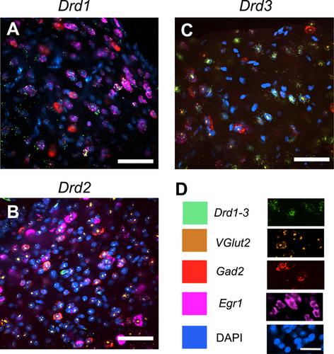
Highly sensitive in situ hybridization procedures (RNAScope) were used to quantify the expression of three dopamine receptors (Drd1, Drd2, and Drd3) in two song control nuclei (HVC and the Area X of the basal ganglia) that are known to receive dopaminergic inputs and in the periaqueductal gray (PAG) of male and female canaries. Both sexes were treated with testosterone to ensure they would sing actively. We also determined the excitatory versus inhibitory phenotype of the cells expressing these receptors as well as their activation following a period of song production. The three receptor types were identified in each brain area, with the exception of Drd3 in Area X. The density of cells expressing each receptor varied as a function of receptor type and brain area. Surprisingly few sex differences were detected; they do not seem to explain the sex differences in testosterone-induced song. Overall, the density of Drd-positive cells was much lower in PAG than in the two song control nuclei. In HVC, the majority of cells expressing the three receptor subtypes were VGlut2-positive, whereas colocalization with Vglut2 occurred in few cells in Area X and in an intermediate proportion of cells in PAG. The number of inhibitory cells expressing dopamine receptors was limited. Most dopaminoceptive cells in Area X did not express either excitatory or inhibitory markers. Finally, cellular activation during singing behavior, as measured by the expression of Egr1, was observed in cells expressing each of the three dopamine receptor subtypes, except Drd3 in the PAG.
期刊介绍:
Established in 1891, JCN is the oldest continually published basic neuroscience journal. Historically, as the name suggests, the journal focused on a comparison among species to uncover the intricacies of how the brain functions. In modern times, this research is called systems neuroscience where animal models are used to mimic core cognitive processes with the ultimate goal of understanding neural circuits and connections that give rise to behavioral patterns and different neural states.
Research published in JCN covers all species from invertebrates to humans, and the reports inform the readers about the function and organization of nervous systems in species with an emphasis on the way that species adaptations inform about the function or organization of the nervous systems, rather than on their evolution per se.
JCN publishes primary research articles and critical commentaries and review-type articles offering expert insight in to cutting edge research in the field of systems neuroscience; a complete list of contribution types is given in the Author Guidelines. For primary research contributions, only full-length investigative reports are desired; the journal does not accept short communications.

 求助内容:
求助内容: 应助结果提醒方式:


