Predicting liver ablation volumes with real-time MRI thermometry

IF 9.5 1区 医学 Q1 GASTROENTEROLOGY & HEPATOLOGY
Osman Öcal , Olaf Dietrich , Sergio Lentini , Pierre Bour , Thibaut Faller , Valery Ozenne , Florian Maier , Matthias Philipp Fabritius , Daniel Puhr-Westerheide , Vanessa F. Schmidt , Elif Öcal , Ricarda Seidensticker , Moritz Wildgruber , Jens Ricke , Max Seidensticker
{"title":"Predicting liver ablation volumes with real-time MRI thermometry","authors":"Osman Öcal ,&nbsp;Olaf Dietrich ,&nbsp;Sergio Lentini ,&nbsp;Pierre Bour ,&nbsp;Thibaut Faller ,&nbsp;Valery Ozenne ,&nbsp;Florian Maier ,&nbsp;Matthias Philipp Fabritius ,&nbsp;Daniel Puhr-Westerheide ,&nbsp;Vanessa F. Schmidt ,&nbsp;Elif Öcal ,&nbsp;Ricarda Seidensticker ,&nbsp;Moritz Wildgruber ,&nbsp;Jens Ricke ,&nbsp;Max Seidensticker","doi":"10.1016/j.jhepr.2024.101199","DOIUrl":null,"url":null,"abstract":"<div><h3>Background &amp; Aims</h3><div>MRI guidance offers better lesion targeting for microwave ablation of liver lesions with higher soft-tissue contrast, as well as the possibility of real-time thermometry. This study aims to evaluate the correlation of real-time MR thermometry-predicted lesion volume with the ablation zone in postprocedural first-day images.</div></div><div><h3>Methods</h3><div>This single-center retrospective analysis evaluated prospectively included patients who underwent MRI-guided microwave ablation with real-time thermometry between December 2020 and July 2023. All procedures were performed under general anesthesia on a 1.5 T MRI scanner. Real-time thermometry data were acquired using multi-slice gradient-echo echoplanar imaging sequences, and thermal dose maps (CEM43 of 240 min as a threshold) were created. The volume of tissue exposed to a lethal thermal dose in MR thermometry (thermal dose) was compared with the ablation zone volume in portal phase T1w MRI on the postprocedural first day using the Pearson correlation test, and visual quantitative assessment by radiologists was performed to evaluate the similarity of shapes and volumes.</div></div><div><h3>Results</h3><div>Out of 30 patients with 33 lesions with thermometry images, six (18.1%) lesions were excluded because of artifacts limiting interpretation of thermal dose volume. Twenty-four patients with 27 lesions (20 male, age 63.1 ± 9.1 years) were evaluated for the volume correlation. The volume of thermal dose-predicted lesions and the postprocedural first-day ablation zones showed a strong correlation (R = 0.89, <em>p</em> &lt;0.001). Similarly, visual similarity of molecular resonance thermometry-predicted shape and the ablation zone shape was graded as perfect in 23 (85.1%) lesions.</div></div><div><h3>Conclusions</h3><div>Real-time thermal dose-predicted volumes show very good correlation with the ablation zone volumes in images obtained 1 day after the procedure, which could reduce the local recurrence rates with the possibility of re-ablating lesions within the same procedure.</div></div><div><h3>Impact and implications:</h3><div>Heat-based ablation is an established treatment for liver tumors; however, there is a considerable rate of incomplete treatment because of the lack of real-time visualization of the treated area during treatment. Our results show that MRI-guided ablation enables the visualization of the treatment area in real-time with high accuracy using a special technique of MR thermometry in patients with liver tumors.</div></div>","PeriodicalId":14764,"journal":{"name":"JHEP Reports","volume":null,"pages":null},"PeriodicalIF":9.5000,"publicationDate":"2024-08-31","publicationTypes":"Journal Article","fieldsOfStudy":null,"isOpenAccess":false,"openAccessPdf":"","citationCount":"0","resultStr":null,"platform":"Semanticscholar","paperid":null,"PeriodicalName":"JHEP Reports","FirstCategoryId":"3","ListUrlMain":"https://www.sciencedirect.com/science/article/pii/S2589555924002039","RegionNum":1,"RegionCategory":"医学","ArticlePicture":[],"TitleCN":null,"AbstractTextCN":null,"PMCID":null,"EPubDate":"","PubModel":"","JCR":"Q1","JCRName":"GASTROENTEROLOGY & HEPATOLOGY","Score":null,"Total":0}
引用次数: 0

Abstract

Background & Aims

MRI guidance offers better lesion targeting for microwave ablation of liver lesions with higher soft-tissue contrast, as well as the possibility of real-time thermometry. This study aims to evaluate the correlation of real-time MR thermometry-predicted lesion volume with the ablation zone in postprocedural first-day images.

Methods

This single-center retrospective analysis evaluated prospectively included patients who underwent MRI-guided microwave ablation with real-time thermometry between December 2020 and July 2023. All procedures were performed under general anesthesia on a 1.5 T MRI scanner. Real-time thermometry data were acquired using multi-slice gradient-echo echoplanar imaging sequences, and thermal dose maps (CEM43 of 240 min as a threshold) were created. The volume of tissue exposed to a lethal thermal dose in MR thermometry (thermal dose) was compared with the ablation zone volume in portal phase T1w MRI on the postprocedural first day using the Pearson correlation test, and visual quantitative assessment by radiologists was performed to evaluate the similarity of shapes and volumes.

Results

Out of 30 patients with 33 lesions with thermometry images, six (18.1%) lesions were excluded because of artifacts limiting interpretation of thermal dose volume. Twenty-four patients with 27 lesions (20 male, age 63.1 ± 9.1 years) were evaluated for the volume correlation. The volume of thermal dose-predicted lesions and the postprocedural first-day ablation zones showed a strong correlation (R = 0.89, p <0.001). Similarly, visual similarity of molecular resonance thermometry-predicted shape and the ablation zone shape was graded as perfect in 23 (85.1%) lesions.

Conclusions

Real-time thermal dose-predicted volumes show very good correlation with the ablation zone volumes in images obtained 1 day after the procedure, which could reduce the local recurrence rates with the possibility of re-ablating lesions within the same procedure.

Impact and implications:

Heat-based ablation is an established treatment for liver tumors; however, there is a considerable rate of incomplete treatment because of the lack of real-time visualization of the treated area during treatment. Our results show that MRI-guided ablation enables the visualization of the treatment area in real-time with high accuracy using a special technique of MR thermometry in patients with liver tumors.
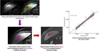
Abstract Image

利用实时磁共振成像测温技术预测肝脏消融体积
背景& 目的MRI引导为肝脏病变的微波消融提供了更好的病灶定位,软组织对比度更高,而且可以进行实时测温。本研究旨在评估实时磁共振温度计预测的病灶体积与术后第一天图像中消融区的相关性。方法这项单中心回顾性分析评估了 2020 年 12 月至 2023 年 7 月期间在磁共振引导下接受实时温度计微波消融术的前瞻性纳入患者。所有手术均在 1.5 T 核磁共振扫描仪上全身麻醉下进行。使用多切片梯度回波回旋成像序列获取实时测温数据,并绘制热剂量图(以240分钟的CEM43为阈值)。使用皮尔逊相关性检验将 MR 测温中暴露于致死热剂量的组织体积(热剂量)与术后第一天门相 T1w MRI 中的消融区体积进行比较,并由放射科医生进行视觉定量评估,以评价形状和体积的相似性。对 24 名患者的 27 个病灶(20 名男性,年龄为 63.1 ± 9.1 岁)进行了体积相关性评估。热剂量预测的病灶体积与手术后第一天的消融区显示出很强的相关性(R = 0.89,p <0.001)。同样,在 23 个(85.1%)病灶中,分子共振测温预测形状与消融区形状的视觉相似度被评为完美。结论实时热剂量预测体积与术后 1 天获得的图像中的消融区体积显示出很好的相关性,这可以降低局部复发率,并有可能在同一次手术中再次消融病灶。影响和意义:热消融是一种治疗肝脏肿瘤的成熟方法,但由于治疗过程中缺乏对治疗区域的实时观察,因此存在相当高的治疗不完全率。我们的研究结果表明,磁共振成像引导下的消融术能利用磁共振测温这一特殊技术实时、高精度地观察肝肿瘤患者的治疗区域。
本文章由计算机程序翻译,如有差异,请以英文原文为准。
求助全文
约1分钟内获得全文 求助全文
来源期刊
JHEP Reports
JHEP Reports GASTROENTEROLOGY & HEPATOLOGY-
CiteScore
12.40
自引率
2.40%
发文量
161
审稿时长
36 days
期刊介绍: JHEP Reports is an open access journal that is affiliated with the European Association for the Study of the Liver (EASL). It serves as a companion journal to the highly respected Journal of Hepatology. The primary objective of JHEP Reports is to publish original papers and reviews that contribute to the advancement of knowledge in the field of liver diseases. The journal covers a wide range of topics, including basic, translational, and clinical research. It also focuses on global issues in hepatology, with particular emphasis on areas such as clinical trials, novel diagnostics, precision medicine and therapeutics, cancer research, cellular and molecular studies, artificial intelligence, microbiome research, epidemiology, and cutting-edge technologies. In summary, JHEP Reports is dedicated to promoting scientific discoveries and innovations in liver diseases through the publication of high-quality research papers and reviews covering various aspects of hepatology.
×
引用
GB/T 7714-2015
复制
MLA
复制
APA
复制
导出至
BibTeX EndNote RefMan NoteFirst NoteExpress
×
提示
您的信息不完整,为了账户安全,请先补充。
现在去补充
×
提示
您因"违规操作"
具体请查看互助需知
我知道了
×
提示
确定
请完成安全验证×
copy
已复制链接
快去分享给好友吧!
我知道了
右上角分享
点击右上角分享
0
联系我们:info@booksci.cn Book学术提供免费学术资源搜索服务,方便国内外学者检索中英文文献。致力于提供最便捷和优质的服务体验。 Copyright © 2023 布克学术 All rights reserved.
京ICP备2023020795号-1
ghs 京公网安备 11010802042870号
Book学术文献互助
Book学术文献互助群
群 号:481959085
Book学术官方微信