{"title":"Myosin VI Is Associated With the Endoplasmic Reticulum in Regions of Sertoli Cells Containing Tubulobulbar Complexes","authors":"Samuel Tretjakov, Prunveer Palia, A. Wayne Vogl","doi":"10.1002/cm.21949","DOIUrl":null,"url":null,"abstract":"<p>Myosin VI has been reported by others to localize in association with various regions of apical tubulobulbar complexes (TBCs) at sites of attachment between Sertoli cells and late spermatids in the mouse. Tubulobulbar complexes internalize “intact” intercellular junctions during sperm release and during spermatocyte translocation through the blood-testis barrier. Here, we use super-resolution (STED—stimulated emission depletion) and electron microscopy of immunolabeled sections of rat testis to clearly define the localization of anti-myosin VI reactivity both at apical and basal sites in the epithelium. In data stacks collected by STED imaging, staining at TBCs was predominantly associated with bulb regions of the complexes. At apical sites, when data stacks were analyzed with an Imaris software, staining appeared around and extended between adjacent bulbs. At basal sites, in addition to labeling at TBC bulbs, reactive sites appeared concentrated in regions close to but not directly associated with intercellular junctions. At the ultrastructural level, labeling was predominantly associated with cisternae of the endoplasmic reticulum associated with the bulbs of TBCs and near to basal junction complexes. We conclude that myosin VI may be associated with specific subdomains of the endoplasmic reticulum related to TBC bulbs and associated basal junction complexes between Sertoli cells.</p>","PeriodicalId":55186,"journal":{"name":"Cytoskeleton","volume":"82 6","pages":"333-343"},"PeriodicalIF":1.6000,"publicationDate":"2024-10-10","publicationTypes":"Journal Article","fieldsOfStudy":null,"isOpenAccess":false,"openAccessPdf":"https://onlinelibrary.wiley.com/doi/epdf/10.1002/cm.21949","citationCount":"0","resultStr":null,"platform":"Semanticscholar","paperid":null,"PeriodicalName":"Cytoskeleton","FirstCategoryId":"99","ListUrlMain":"https://onlinelibrary.wiley.com/doi/10.1002/cm.21949","RegionNum":4,"RegionCategory":"生物学","ArticlePicture":[],"TitleCN":null,"AbstractTextCN":null,"PMCID":null,"EPubDate":"","PubModel":"","JCR":"Q4","JCRName":"CELL BIOLOGY","Score":null,"Total":0}
引用次数: 0
Abstract
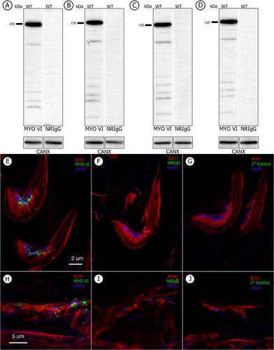
Myosin VI has been reported by others to localize in association with various regions of apical tubulobulbar complexes (TBCs) at sites of attachment between Sertoli cells and late spermatids in the mouse. Tubulobulbar complexes internalize “intact” intercellular junctions during sperm release and during spermatocyte translocation through the blood-testis barrier. Here, we use super-resolution (STED—stimulated emission depletion) and electron microscopy of immunolabeled sections of rat testis to clearly define the localization of anti-myosin VI reactivity both at apical and basal sites in the epithelium. In data stacks collected by STED imaging, staining at TBCs was predominantly associated with bulb regions of the complexes. At apical sites, when data stacks were analyzed with an Imaris software, staining appeared around and extended between adjacent bulbs. At basal sites, in addition to labeling at TBC bulbs, reactive sites appeared concentrated in regions close to but not directly associated with intercellular junctions. At the ultrastructural level, labeling was predominantly associated with cisternae of the endoplasmic reticulum associated with the bulbs of TBCs and near to basal junction complexes. We conclude that myosin VI may be associated with specific subdomains of the endoplasmic reticulum related to TBC bulbs and associated basal junction complexes between Sertoli cells.
期刊介绍:
Cytoskeleton focuses on all aspects of cytoskeletal research in healthy and diseased states, spanning genetic and cell biological observations, biochemical, biophysical and structural studies, mathematical modeling and theory. This includes, but is certainly not limited to, classic polymer systems of eukaryotic cells and their structural sites of attachment on membranes and organelles, as well as the bacterial cytoskeleton, the nucleoskeleton, and uncoventional polymer systems with structural/organizational roles. Cytoskeleton is published in 12 issues annually, and special issues will be dedicated to especially-active or newly-emerging areas of cytoskeletal research.
 求助内容:
求助内容: 应助结果提醒方式:


