Katia Montanha-Andrade, Paula Paes Ferreira, Ana Carolina Velasco Pondé DE Sena, Patricia R Cury, Ieda M Crusoé-Rebello
{"title":"Tomographic diagnosis of alveolar bone coverage impact in orthodontic planning: cross-sectional study.","authors":"Katia Montanha-Andrade, Paula Paes Ferreira, Ana Carolina Velasco Pondé DE Sena, Patricia R Cury, Ieda M Crusoé-Rebello","doi":"10.1590/2177-6709.29.5.e242446.oar","DOIUrl":null,"url":null,"abstract":"<p><strong>Introduction: </strong>Alveolar bone coverage can be diagnosed through cone beam computed tomography (CBCT) and this information can prevent orthodontic tooth movement beyond the biological limit.</p><p><strong>Objective: </strong>This study evaluated the impact of the bone coverage (BC) diagnosis by CBCT in the orthodontists' planning.</p><p><strong>Methods: </strong>One hundred fifty-nine Brazilian orthodontists suggested treatment plans for six patients at two different times, using two sequential questionnaires. The first questionnaire consisted of extra and intra-oral photographs, one panoramic radiograph; one lateral cephalometric radiograph with Steiner and Tweed analysis, and the patient chief complaint. The second questionnaire included the same presentations of cases with tomographic images and the radiologist's report. The McNemar test assessed the difference between the first and the second treatment plans.</p><p><strong>Results: </strong>In all six cases, most participants changed the treatment plan after evaluating the CBCT images and the radiologist's report (93.7% in case 5, 78.6% in case 4, 74.2% in case 3, 69.8% in case 6, 66% in case 2 and 61% in case 1; p≤0.01).</p><p><strong>Conclusion: </strong>The evaluation of bone coverage through CBCT images has a substantial impact on the orthodontic diagnosis and planning of the Brazilian orthodontists.</p>","PeriodicalId":38720,"journal":{"name":"Dental Press Journal of Orthodontics","volume":"29 5","pages":"e242446"},"PeriodicalIF":0.0000,"publicationDate":"2024-10-07","publicationTypes":"Journal Article","fieldsOfStudy":null,"isOpenAccess":false,"openAccessPdf":"https://www.ncbi.nlm.nih.gov/pmc/articles/PMC11457962/pdf/","citationCount":"0","resultStr":null,"platform":"Semanticscholar","paperid":null,"PeriodicalName":"Dental Press Journal of Orthodontics","FirstCategoryId":"1085","ListUrlMain":"https://doi.org/10.1590/2177-6709.29.5.e242446.oar","RegionNum":0,"RegionCategory":null,"ArticlePicture":[],"TitleCN":null,"AbstractTextCN":null,"PMCID":null,"EPubDate":"2024/1/1 0:00:00","PubModel":"eCollection","JCR":"Q2","JCRName":"Medicine","Score":null,"Total":0}
引用次数: 0
Abstract
Introduction: Alveolar bone coverage can be diagnosed through cone beam computed tomography (CBCT) and this information can prevent orthodontic tooth movement beyond the biological limit.
Objective: This study evaluated the impact of the bone coverage (BC) diagnosis by CBCT in the orthodontists' planning.
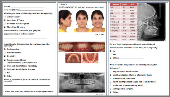
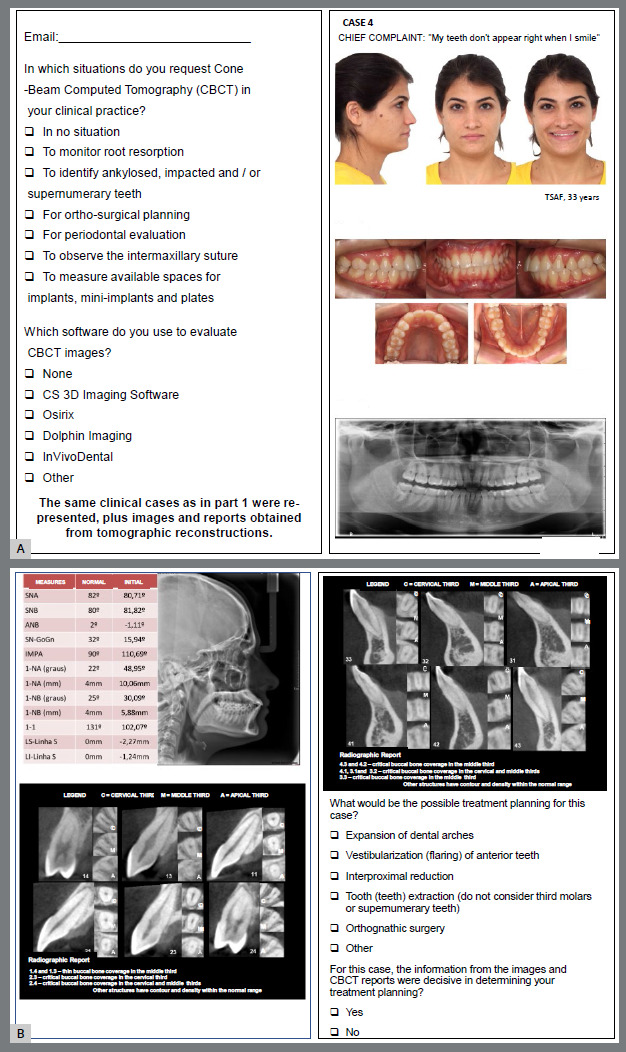
Methods: One hundred fifty-nine Brazilian orthodontists suggested treatment plans for six patients at two different times, using two sequential questionnaires. The first questionnaire consisted of extra and intra-oral photographs, one panoramic radiograph; one lateral cephalometric radiograph with Steiner and Tweed analysis, and the patient chief complaint. The second questionnaire included the same presentations of cases with tomographic images and the radiologist's report. The McNemar test assessed the difference between the first and the second treatment plans.
Results: In all six cases, most participants changed the treatment plan after evaluating the CBCT images and the radiologist's report (93.7% in case 5, 78.6% in case 4, 74.2% in case 3, 69.8% in case 6, 66% in case 2 and 61% in case 1; p≤0.01).
Conclusion: The evaluation of bone coverage through CBCT images has a substantial impact on the orthodontic diagnosis and planning of the Brazilian orthodontists.
期刊介绍:
The Dental Press Journal of Orthodontics publishes scientific research articles, significant reviews, clinical and technical case reports, brief communications, and other materials related to Orthodontics and Facial Orthopedics.

 求助内容:
求助内容: 应助结果提醒方式:


